Download
1 / 165
1.7k likes | 2.23k Views
Chapter 9 Function of the Sense Organs. Introduction. Human life would be very different without the ability to sense and perceive external stimuli Imagine your world without the ability to see, hear, smell, touch, and feel.

E N D
Chapter 9 Function of the Sense Organs
Introduction • Human life would be very different without the ability to sense and perceive external stimuli • Imagine your world without the ability to see, hear, smell, touch, and feel
Environmental sensation is limited to those forms of energy that sensory receptors are designed to detect. • Sensory receptors may convey information to the cortex with awareness or perception and may lead to cerebrally controlled responses. • Sensory receptors also serve as afferent pathways for reflex action with or without conscious sensation.
Section 1 Physiology of the Receptor and Sense Organs I Concept and Classification of the Receptor and Sense Organs
Sensory Receptors • Receptorsarespecialized nerve cells that transduce energy into neural signals • Receptors lack axons, form synapses with dendrites of other sensory neurons • Receptors are “mode” specific • “Law of Specific Nerve Energies”: sensory messages are carried on separate channels to different areas of the brain • Receptors detect a small range of energy levels • Eye: 400-700 nM • Ear: 20-20,000 Hz • Taste buds: specific chemicals
General sensory receptor structure • Free nerve endings: dendrites interspersed among other cells/tissues (pain, temperature, touch)
General sensory receptor structure • Encapsulated nerve endings: dendrites with special supporting structures (mechanoreceptors and proprioceptors)
Classification of Receptors: • Location • Externoceptors • Located on the body surface or specialized to detect external stimuli • Pressure, pain, temp, touch, etc. • 2) Visceroceptors • Located within internal organs, detect internal stimuli • Blood pressure, pain, fullness. • 3) Proprioceptors • Found in the joints and muscles • Also in the vestibular structures and the semicircular canals of the inner ear. Limb and body position and movment.
2 Modalities • Mechanoceptive • Detects stimuli which mechanically deform the receptor; • Pressure, vibration, touch, sound. • 2) Thermoceptive • Detects changes in temperature; hot/cold • 3) Nociceptive (pain) • Detects damage to the structures • 4) Photoreceptors • Detect light; vision, retinal of the eye • 5) Chemoceptive • Detect chemical stimuli; CO2 and O2 in the blood, glucose, small, taste.
3. Complexity • Simple receptors • Usually a single modified dendrite • General sense • Touch, pressure, pain, vibration, temperature • 2) Complexity • High modified dendrites, organized into complex structures; ear, eye. • Special senses: • Vision, hearing, smell, taste
Sensation: Receiving messages • Stimuli: What messages can be received? • Anything capable of exciting a sensory receptor cell can be defined as a “stimulus” • Examples include: sound, light, heat, cold, odor, color, touch, and pressure
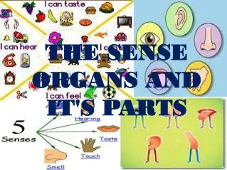
Sensation: Receiving messages about the world • Sense organs operate through sensory receptor cells that receive external forms of energy and translate these external forms into neural impulses that can be transmitted to the brain • There are two types of sense organs which we will examine in this chapter
Basic Function Sequence of Events in a Receptor Reception Receptor Protein Activated Enzyme Cascade (in some cases) Receptor Ion Channels opened (or closed) Receptor Current Transduction Receptor Potential Modulated Transmitter Release from Receptor Cell Modulated Impulse Frequency in Second Order Neuron Modulated Impulse Frequency in Receptor Cell Axon Transmission Stimulus Amplification
Each type of receptor is highly sensitive to one type of stimulus for which it is designed and yet is almost nonresponsive to normal intensities of other type of stimuli. • The stimulus to which a given receptor has the lowest threshold is termed the adequate stimulus of the sensory receptor. • For instance, the roes and cones are highly responsive to light but almost completely nonresponsive to heat and cold.
2.Transduction of Sensory Receptors • Transduction: The process by which an environmental stimulus becomes encoded as a sequence of nerve impulses in an afferent nerve fiber is called sensory transduction • Sense orgrans transduce sensory energy into neural (bioelectrical) energy • Converting one type of energy into another type is the process of transduction • Your brain only deals with bioelectrical impulses so transduction must occur; what cannot be transduced cannot be a stimulus
Principles of Transduction • Different kinds of receptor are activated in different ways but the first stage in sensory transduction is the generation of a graded receptor potential. • The magnitude of the stimulus is related to that of the receptor potential which in turn is related to either • a) the sequence or frequency of all-or-none action potentials generated in the afferent nerve fiber; • b) modulated release of transmitter from the receptor cell generating a sequence of action potentials in a second order neurone.
Receptor/Generator Potential Receptor potentials: Changes in the transmembrane potential of a receptor caused by the stimulus. Generator Potential: A receptor potential that is strong enough (reaches threshold) to generate an action potential. Remember that APs are all-or-none. The stronger the sitmulus (above threshold) the more APs are fired over a given time period; this is translated by the CNS as a strong sensation.
Sensory Adaptation is one form of Integration Phasicreceptors quickly adapt. The frequency of action potentials diminishes or stops if the stimulus is unchanging. Tonic receptors adapt slowly or not at all. Most exteroreceptors(receptors that monitor the external environment) are phasic receptors.
Phasic receptors alert us to changes in sensory stimuli and are in part responsible for the fact that we can cease paying attention to constant stimuli. The slowly adapting receptors (tonic receptors), such as the pain receptors and the baroreceptors of the arterial tree, are useful in situations requiring maintained information about a stimulus.
4. Encoding of Sensory Receptor The quality of the stimulus is encoded in the frequency of the action potentials transmitted down the afferent fibre and the number of sensory receptors activated.
Stretch Receptors: Weak stretch causes low impulse frequency on neuron leaving receptor. Strong stretch causes high impulse frequency on neuron leaving receptor. Frequency Code Membrane potential Time
Summary • The external & internal environments are monitored by sensory receptors. • Each type of receptor is excited most effectively by only one modality of stimulus known as the adequate stimulus. • The stimulus is converted into an electrical potential. • Stimuli are detected as either static or dynamic events. • The intensity & duration of the stimulus is frequency coded as bursts of action potentials in the primary afferent nerve.
Functions of the Complete Eye • Eye functions like a camera • Iris allows light into eye • Cornea, Lens & humors focus light onto retina • Light striking retina is converted into action potentials relayed to brain
Structure of • the Eyeball • A slightly irregular hollow sphere with anterior and posterior poles • The wall is composed of three tunics – fibrous, vascular, and sensory • The internal cavity is filled with fluids called humors • The lens separates the internal cavity into anterior and posterior segments
Anatomy of the Eye • Three coats or tunics • Fibrous: Consists of sclera and cornea • Vascular: Consists of choroid, ciliary body, iris • Nervous: Consists of retina
1. Fibrous Tunic • Forms the outermost coat of the eye and is composed of: • Opaque sclera • Clear cornea • The sclera protects the eye and anchors extrinsic muscles • The cornea lets light enter the eye
2. Vascular Tunic (uvea): • Has three regions: choroid, ciliary body, and iris • Choroid region • A dark brown membrane that forms the posterior portion of the uvea • Supplies blood to all eye tunics
Vascular Tunic: Ciliary Body • A thickened ring of tissue surrounding the lens • Composed of smooth muscle bundles (ciliary muscles) • Anchors the suspensory ligament that holds the lens in place
Vascular Tunic: Iris • The colored part of the eye • Pupil – central opening of the iris • Regulates the amount of light entering the eye during: • Close vision and bright light – pupils constrict • Distant vision and dim light – pupils dilate
Pupil Dilation and Constriction Figure 15.9
3. The Retina and its Parts Optic Nerve, Blind Spot, Fovea
The Retina and its Parts • Retina:inner layer on back of eye that contains “light-sensitive” rods and cones • Optic Nerve: bundle of axons running from retina to visual (occipital) cortex • Blind Spot:spot on the retina where optic nerve exits eye, there are no receptors (rods or cones) there • Fovea:center of the retina where “acuity” (ability to see fine detail) is greatest
II The Image-Forming Mechanism The images of objects in the environment are focused on the retina.
1. Principle of Optics Light rays are bent (refracted) when they pass from one medium into a medium of a different density. Parallel light rays striking a biconvex lens are refracted to a point (principal focus) behind the lens. The principle focus is on a line passing through the centers of a curvature of the lens, at the principal focal distance.
For practical purpose, light rays from an object that strike a lens more than 20 ft (6 m) away are considered to be parallel. The rays from an object closer than 20 ft are diverging and are therefore brought to a focus farther back on the principal axis than the principal focus. Biconcave lenses cause light rays to diverge.
2.Optic Characteristics of Refractive System in Human Eye The refractive system of the human eye is composed of the cornea, aqueous humor, crystalline lens, and vitreous humor. When light coming from an object is brought to a focus, an image is formed. In the normal human eye, parallel rays of light entering the eye are focused to an image just on the retina. This ideal condition is called emmertropia.
So the image of distant object (6 m away) will be focused on the retina in the emmertropic eye.
3. Accommodation When the ciliary muscle is relaxed, parallel light rays striking the optically normal (emmetropic) eyes are brought to a focus on the retina. As long as this relaxation is maintained, rays from objects closer than 6 m from the observer are brought to a focus behind the retina, and consequently the objects appear blurred.
This problem can be solved by increasing the curvature or refractive power of the lens. The process whereby near objects are brought to a sharp focus on the retina is called accommodation of eye or visual accommodation. Accommodation involves following reflexes.
(1) Accommodation of lens • Increase bulging (refraction) of lens • Via contraction of ciliary muscle, relaxes the suspensory ligaments (parasympathetic fibers)
Accommodation of Lens. The solid lines represent the shape of the lens, iris, and ciliary body at rest, and the dotted lines represent the shape during accommodation.