Download
1 / 68
840 likes | 3.04k Views
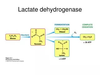
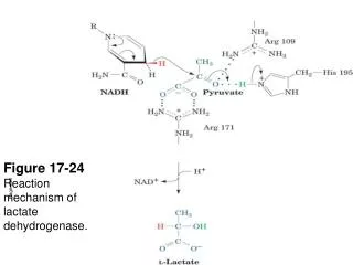
1. It is an enzyme which catalyzes the reactionLactate ?? Pyruvate 2. This reaction helps the re-oxidation of NADH, H into NAD 3. It has 5 isoenzymes: LD1, LD2, LD3, LD4 and LD5.4. Medical importance:5. Estimation of the activity of lactate dehydrogenase enzyme helps in the diagnosis of heart and liver diseases:a) LD1: Elevated in some heart diseases e.g. myocardial infraction.b) LD5: Elevated in some liver diseases as acute viral hepatitis. .

E N D
1. Lactate Dehydrogenase Prof. Dr. Laila Fadda
2. 1. It is an enzyme which catalyzes the reaction
Lactate ?? Pyruvate
2. This reaction helps the re-oxidation of NADH, H+ into NAD+
3. It has 5 isoenzymes: LD1, LD2, LD3, LD4 and LD5.
4. Medical importance:
5. Estimation of the activity of lactate dehydrogenase enzyme helps in the diagnosis of heart and liver diseases:
a) LD1: Elevated in some heart diseases e.g. myocardial
infraction.
b) LD5: Elevated in some liver diseases as acute viral hepatitis.
3. In vitro inhibition of glycolvsis:
Arsenate: by competing with Pi in the reaction:
Glyceraldhyde-3-p 1,3 diphosphoglycerate
lodoacetate: by inhibiting glyceraldhyde-3-p dehydrogenase.
Fluoride: Inhibits enolase enzyme. Clinical laboratories use fluoride to inhibit glycolysis by adding it to the blood before measuring blood glucose.
4. Fermentation:
Definition: This is conversion of glucose into ethanol by yeast enzymes.
Pyruvate is formed by the same series of reactions of glycolysis.
Then pyruvate is converted into acetaldehyde, then ethanol as follows.
Thus the end product of fermentation is CO2 ethanol.
6. Regulation of glycolvsis:
The rate of glycolysis is regulated by controlling of the 3 irreversible enzymes (key enzymes). These enzymes catalyze what is called committed reactions of the pathway. These enzymes are glucokinase (hexokinase), phospho-fructokinase-1 and pyruvate kinase.
7. 1. Hormonal regulation:
a) Insulin: Stimulates synthesis of all key enzymes of glycolysis. It is secreted after meal (in response to high blood glucose level).
b) Glycogen: Inhibits the activity of all key enzymes of glycolysis. It is secreted in response to low blood glucose level.
2. Energy regulation:
a) High level of ATP inhibits PFK-1 and pyruvate kinase.
b) High level of ADP and AMP stimulate PFK-1.
8. 3. Substrate regulation:
a) Glucose-6-phosphate inhibits hexokinase (and not glucokinase).
b) Fructose 2,6 bisphosphate stimulates phosphofructokinase-1.
c) Citrate inhibits phosphofructokinase-1.
d) Fructose 1,6 bisphosphate stimulates pyruvate kinase.
9. Lactate:
Sources and fate of lactate:
a) Sources:
From glycolysis especially in RBCs due to absence of mitochondria and muscle during exercises due to oxygen lack.
10. b) Fate:
1) Glucose formation: [through lactic acid (Cori cycle)]:
Lactate formed in muscles and RBCs may be diffuse to blood then to the liver.
In the liver, lactate is converted to glucose by gluconeogenesis. Glucose may diffuse back to the blood, then to red cells or muscles to be used for production of energy. This cycle is called: Lactic acid or Cori cycle.
Definition of Cori cycle: It is the conversion of glucose to lactate in peripheral tissues, followed by conversion of lactate into glucose in liver.
11. 2) Conversion into pyruvate: If oxygen gets available, lactate is converted into pyruvate, which proceeds into Krebs' cycle.
3) Lactate may be accumulated in muscles causing muscle fatigue.
4) Lactate may be excreted in urine and sweat.
c) Blood level of lactate:
Normal blood lactate level is: 1-2 mmol/L.
12. Clinical aspects of glycolvsis:
1. There are many diseases associated with impaired glycolysis. They include:
1. Pyruvate kinase deficiency.
2. Hexokinase deficiency.
3. Lactic acidosis.
2) Pyruvate kinase (PK) deficiency:
a) This leads to excessive hemolysis of RBCs leading to hemolytic anemia.
b) Genetic deficiency of PK enzyme causes decrease the rate of glycolysis and decrease production of ATP.
c) ATP is required for Na' -K' ATPase, which is important for stability of RBCs.
13. 3. Hexokinase deficiency:
Leads to hemolytic anemia due to decrease ATP production. The mechanism is similar to that of PK deficiency.
4. Lactic acidosis:
a) Definition and mechanism of lactic acidosis:
1) It is the lowered blood pH and bicarbonate levels due to increased blood lactate above normal level.
OH OH
CH3-C -- COOH + NaHCO3 CH3-C-COONa + H2CO3 CO2 + H2O
14. Lactate Sodium bicarbonate Sodium lactate Carbonic acid
2) This depletes bicarbonate ? pH of blood Lactic acidosis which if severe may lead to coma.
b) Causes of lactic acidosis:
It results from increased formation or decreased utilization of lactate.
1) Increased formation of lactate as in severe muscular exercises.
15. 2) Decreased utilization of lactate in tissues: it occurs in cases of anoxia or lack of oxygen. This is because oxygen is essential for conversion of lactate into pyruvate, which proceeds into acetyl CoA, and Krebs' cycle.
Tissue anoxia may occur in conditions that impair blood flow e.g. myocardial infraction, angina pectoris, respiratory disorders, and anemia.
3) Phenformin (cidophage): is oral hypoglycemic, causing excessive anaerobic oxidation of glucose and excess lactate production.
16. II) Mitochondrial pathway for glucose oxidation:
A. Introduction: Complete oxidation of glucose occurs in both cytoplasm (glycolysis) and mitochondria (Krebs' cycle) In the presence of O2 pyruvate (the product of glycolysis) passes by special pyruvate transporter into mitochondria which proceeds as follows:
1. Oxidative decarboxylation of pyruvate into acetyl CoA.
2. Acetyl CoA is then oxidized completely to CO2, H2O through Krebs' cycle.
17. B. Oxidative decarboxylation of pvruvate to acetvl coenzvme A (= acetyl CoA):
1. Enzyme: Pyruvate dehydrogenase (PHD) complex.
a) This enzyme complex contains 4 subunits, which catalyze the reaction in 4 steps.
18. b) This enzyme needs 5 coenzymes (all are vitamin B complex derivatives):
1) Vitamin B1 = Thiamin diphosphate = TPP.
2) Lipoic acid = L -SH.-SH (reduced), L-s-s (oxidized).
3) Coenzyme A = CoASH.
4) Flavin adenine dinucleotide = FAD.
5) Nicotinamide adenine dinucleotide = NAD+
19. c) Location: PDH is located within the mitochondrial matrix.
1) Energy production: (3ATP):
Oxidative decarboxylation of pyruvate to acetyl CoA produces one molecule of NADH,H+. This produces 3 ATP molecules through respiratory chain phosphorylation
20. 2) Regulation of oxidative decarboxylation (PHD):
21. a) Factors stimulating [+] PHD:
1) Pyruvate.
2) CoASH.
3) NAD+
4) Insulin hormone.
b) Factors inhibiting [-] PHD:
1) NADH,H+
2) ATP.
3) Acetyl CoA.
4) Calcium ions.
22. c) Mechanism of regulation:
1) PDH exists in two forms:
Phosphorylated (inactive), dephosphorylated (active).
2) Protein kinase enzyme converts active into inactive enzyme.
3) Phosphatase enzyme converts inactive into active.
3) Invitro inhibition of PDH:
a) Arsenic.
b) Thiamin (6,) deficiency.
23. Kreb's cycle: [also known as citric acid cycle (CAC) or tricarboxylic acid cycle (TCA) or catabolism of acetyl CoA]:
1. Definition: TCA is a series of reactions in which acetyl CoA is oxidized into C2O1 H2O and energy.
2. Location: Mitochondria.
25. 3. Steps:
a) The enzymes of TCA cycle are present in the mitochondrial matrix either free or attached to the inner surface of the mitochondrial membrane.
b) The cycle is started by acetyl CoA (2 carbons) and oxaloacetate (4 carbons) to form citrate (6 carbons). It ends by oxaloacetate (4 carbons). The difference between the starting compound (6 carbons) and the ending compound (4 carbons) is 2 carbons that are removed in the form of 2 CO2. These 2 carbons are derived from acetyl CoA. For this reasons acetyl CoA is completely catabolized in TCA and never gives glucose.
26. 4. Energy production of TCA: (Energy catabolism of acetyl CoA):
a) Oxidation of one molecule of acetyl CoA in TCA produces 12 ATP molecules, 11 by respiratory chain phosphorylation and 1 by Substrate level phosphorylation as follows:
28. b) Energy production of complete oxidation of one molecule of glucose:
Glucose oxidation 36 or 38 ATP
Pyruvate oxidation 15ATP.
Acetyl CoA 12 ATP
29. 5. Oxidative decarboxylation of -ketoglutarate to succinyl CoA
It is similar to the conversion of pyruvate to acetyl CoA.
a) Enzymes: ketoglutarate dehydrogenase complex.
b) Coenzymes: 5: TPP, Lipoic acid, CoASH, FAD and NAD+.
6. Functions (significance) of TCA:
The cycle is amphibolic i.e. it has catabolic (breakdown) and anabolic (formation) functions.
30. Energy: 12 ATP
Catabolic functions: Oxidation of carbohydrate, lipids and proteins
Anabolic function: Formation of:
Amino acids
Glucose
Heme
Fatty acid and cholesterol
CO2 and over-controlling in his relation with the patient.
31. a) Production of energy (12 ATP).
b) Catabolic functions: TCA is the final common pathway for oxidation of carbohydrates, fats and proteins (amino acids).
c) Anabolic functions: Formation of:
1) Amino acids:
-Ketoglutarate Transamination Glutamate.
Oxaloacetate Transamination Aspratate.
32. 2) Glucose: e.g.
Ketoglutarate Gluconeogenesis Glucose.
3) Heme synthesis:
Succinyl CoA Heme.
4) Fatty acid and cholesterol:
Citrate (diffuse to cytoplasm) ? Oxaloacetate + Acetyl
CoA ? Fatty acid and cholesterol.
33. 5) CO2 produced is used in the following (CO2 fixation) reactions:
Pyruvate + CO2 Oxaloacetate Gluconeogenesis Glucose.
Acetyl COA + CO2 Malonyl CoA Fatty acids.
Ammonia + ATP +CO2 Carbamoyl phosphate Urea and pyrimidine.
Propionyl CoA + CO2 Methyl malonyl CoA Odd number fatty acid.
Formation of C6 of purine.
Synthesis of H2CO3 / HCO, buffer.
34. 7. In vitro inhibition of TCA cycle:
a) Flouroacetate (F1-CH2-COSCoA): inhibits aconitase enzyme.
b) Arsenate: inhibits a-ketoglutarate dehydrogenase enzyme.
c) Malonic acid: inhibits succinate dehydrogenase enzyme (competitive inhibition).
35. 8. Regulation of citric acid cycle:
The key enzymes are citrate synthase dehydrogenase and -ketoglutarate dehydrogenase:
a) Citrate synthase:
1) Stimulated by acetyl CoA, oxaloacetate, ADP and NAD�+.
2 ) Inhibited by long chain acyl CoA, citrate, succinyl CoA, ATP and NADH, H+
36. b) Isocitrate dehydrogenase and -ketoglutarate dehydrogenase:
1) Stimulated by NAD+, ADP.
2) Inhibited by NADH, H' and ATP
c) Availability of Oxygen: Citric acid cycle needs oxygen to proceed (i.e. aerobic pathway). This is because in absence of oxygen respiratory chain is inhibited leading to increase NADH,H+ / NAD. NADH, H+ will inhibit TCA cycle.
37. 9. Sources and fate of oxaloacetate:
a) Sources of oxaloacetate:
1) Oxidation of malate: Final step in TCA cycle.
2) Transamination of aspartate: See protein metabolism.
3) Carboxylation of pyruvate: By pyruvate carboxylase and biotin (see gluconeogenesis).
38. 4) Cleavage of citrate:
39. b) Fates of oxaloacetate:
1) Formation of citrate: By citrate synthase (first step in TCA cycle).
2) Reduction to malate.
3) Transamination into aspartic acid.
40. 10. Energy production at substrate level in glucose oxidation:
a) The removal of hydrogen atoms from a compound is accompanied by a release of energy. If this energy is captured in phosphate or sulfate bonds, it will produce high-energy compounds.
b) The high energy compounds formed by glucose oxidation are:
1) Glyceraldhyde-3-p 1,3 BPG (phosphate bond).
2) Phosphoglycerate phosphoenol pyruvate (phosphate bond).
3) Pyruvate Acetyl CoA (sulfate bond(.
4) -Ketoglutarate Succinyl CoA (sulfate bond).
41. 11. Pasteur effect:
a) It is the inhibition of glycolysis (anaerobic oxidation) by the presence of oxygen.
b) Explanation: Aerobic oxidation of glucose produces increased amount of ATP and citrate Those inhibit phosphofructokinase1 (one of the key enzymes of glycolysis) Inhibition of glycolysis.
42. III. Pentose phosphate pathway (Hexose phosphate pathway):
A. Definition: It is an alternative pathway for glucose oxidation where:
1. ATP (energy) is neither produced nor utilized.
2. Its main function is to produced NADPH, H+ and pentoses.
B. Location:
1. Intracellular location: cytoplasm.
43. 2. Organ location: It is active in tissues where NADPH, H+ is needed for fatty acids or steroids synthesis.
a) Adipose tissue and liver: It supplies NADPH, H+ for Lipogenesis
b) Adrenal cortex, ovaries and testis: It supplies NADPH, H' for steroid synthesis.
c) Red cells: It supplies NADPH, H+ for production of glutathione.
d) Retina: It supplies NADPH, H+ for reduction of retinal into retinol.
e) In many tissues: It supplies pentoses for synthesis of nucleotides.
44. C. Reactions (steps):
This pathway occurs in two phases; oxidative and non-oxidative:
1. Oxidative (irreversible) phase: where 3 molecules of glucose are converted into 3 molecules of ribulsose-5- phosphate with production of NADPH, H+ and CO2.
2. Non-oxidative (reversible) phase: Where the 3 molecules of ribulose-5-phosphate are interacted and converted into 2 molecules of glucose-6-phosphate and one molecule of glyceraldhyde-3-phosphate.
45. D. Functions of HMP pathway:
Production of pentoses:
RNA, DNA.
ATP,GTP.. ..etc.
NAD+, FAD. . etc.
Production of NADPH + H+:
Synthesis of substrates e.g. FA, cholesterol. ....etc.
Reduction of glutathione.
Hydroxylation of aromatic compounds.
Phagocytosis and respiratory burst.
46. 1. Production of pentoses:
Which are essential for synthesis of nucleic acids (RNA and DNA), nucleotides (ATP, GTP ....etc) and coenzymes (NAD+, NADP, FAD ... .etc).
47. 2. Production of NADPH, H+ : It is important for
a) Synthesis of many substrates:
1) Synthesis of fatty acids (lipogenesis) cholesterol and other steroid hormones.
2) Synthesis of sphingosine and galacto-lipids.
3) Essential for glucuronic acid metobolism.
4) Synthesis of non essential amino acids.
5) Synthesis of malate from pyruvate by malic enzyme.
48. b) In RBCs: Reduction of glutathione:
49. Reduced glutathione is essential for:
1) Normal integrity of RBCs.
2) Maintenance of SH group of RBCs enzymes.
3) Removal of hydrogen peroxide (H2O), which is a toxic compound that increases cell membrane fragility.
50. c) In liver: Hydroxylation of aromatic and aliphatic compounds:
NADPH, H acts as coenzyme for liver microsomal P-450 monooxygenase system (enzyme). This is the major pathway for the hydroxylation of toxic aromatic and aliphatic compounds such as steroids, alcohols and many drugs converting them into nontoxic compounds.
51. E. Regulation of HMP shunt:
Glucose-6- phosphate dehyrogenase is the key enzyme of HMP-shunt. It is stimulated by insulin and NADP+ and inhibited by NADPH, H+ and acetyl CoA. .
53. F. Differences between HMP shunt and Qlvcolvsis:
54. G. HMP shunt in skeletal muscles:
1. Skeletal muscles are poor in glucose-6-phosphate dehydrogenase enzyme.
2. Skeletal muscles obtain their pentose requirement by reversible reactions of HMP pathway, using fructose-6-phosphate and glyceralde-3-p and the enzymes transketolase and transaldolase.
55. H. Defects of HMP pathway:
Favism (Deficiency of glucose-6-phosphate dehydrogenase enzyme):
1. Definition:
It is a hemolytic anemia (excessive destruction of RBCs) especially after ingestion-of fava beans and some other compounds. It is due to ?G-6-P dehydrogenase enzyme.
56. 2. Mechanism:
a) Deficiency of glucose-6-P dehydrogenase ? Decreased NADPH,H+ which is essential for RBCs to reduce glutathione in the presence of glutathione reductase enzyme.
57. b) Reduced glutathione (G-SH) is needed to remove hydrogen peroxide (H2O2) which is toxic to the cell.
58. c) Deficiency of glucose-6-PDH ? ?NADPH , H+ ??reduced glutathione ? Accumulation of H2O2 ? Hemolysis of RBCs
d) Effect of H2O2 on RBCs:
1) Peroxidation of fatty acids present in cell wall Hemolysis.
2) Conversion of hemoglobin into met-hemoglobin. These toxic compounds increases the red cell membrane fragility.
59. 3. Signs and symptoms of favism:
a) Patients with enzyme deficiency show attacks of hemolytic anemia in the form of severe jaundice and decreased hemoglobin concentration when exposed to 'certain oxidizing agents as:
1) Special food as fava beans.
2) Antimalarial drugs: as primaquine.
3) Antibiotics as streptomycin.
60. 1. Structure of glycogen:
A. Glycogen is homopolysaccharide formed of branched D glucose units (1,4 and 1,6).
B. The primary glycosidic bond is 1-4-linkage
C. Each branch is made of 6-12 glucose units. At the branching point, the chain is attached by 1-6 linkage.
61. II. Location of glycogen: Glycogen is present mainly in cytoplasm of liver and muscles.
A. Liver glycogen is about 120 grams (about 6 % of liver weight).
B. Muscle glycogen is about 350 grams )about 1 % of total muscles weight).
62. III. Functions of glycogen:
A. Liver glycogen: It maintains normal blood glucose concentration especially during the early stage of fast (between meals). After 12-18 hours fasting, liver glycogen is depleted.
B. Muscle glycogen: It acts as a source of energy within the muscle itself especially during muscle contractions.
63. IV. Synthesis of glycogen (glycogenesis):
A. Definition: It is the formation of glycogen in liver and muscles.
B. Substrates for glycogen synthesis:
1. In liver:
a) Blood glucose.
b) Other hexoses: fructose and galactose.
c) Non-carbohydrate sources: (gluconeogenesis) e.g. lactic acid, glycerol and lactate. These are converted first to glucose, then to glycogen.
64. 2. In muscles:
a) Blood glucose only.
C. Steps:
Glucose molecules are the first activated to uridine diphosphate glucose (UDP-G). Then these UDP-G molecules are added to a glycogen primer to form glycogen.
65. 1. Formation of UDP-Glucose (UDP-G):
Note: Glucose is converted into glucose-6-phosphate:
1) In liver: by glucokinase.
2) In muscle: by hexokinase.
66. 2. Formation of glycogen:
UDP-Glucose reacts with glycogen primer, which may be:
a) Few molecules of glucose linked together by a 1-4 linkage.
b) A protein called: glycogenin. UDP-G molecules react with OH of tyrosine of that protein to initiate glycogen synthesis.
c) Glycogen synthase:
By the action of glycogen synthase (key enzyme of glycogenesis), UDP-G molecules are added to glycogen primer causing elongation of the 1-4, branches up to 11 glucose units.
67. 2. Formation of glycogen:
UDP-Glucose reacts with glycogen primer, which may be:
a) Few molecules of glucose linked together by a 1-4 linkage.
b) A protein called: glycogenin. UDP-G molecules react with OH of tyrosine of that protein to initiate glycogen synthesis.
c) Glycogen synthase:
By the action of glycogen synthase (key enzyme of glycogenesis), UDP-G molecules are added to glycogen primer causing elongation of the 1-4, branches up to 11 glucose units.
68. d) Branching enzyme:
It transfers parts of the elongated chains (5-8 glucose residues) to the next chain forming a new a 1-6 glycosidic bond. The new branches are elongated by the glycogen synthase and the process is repeated.
69. V. Breakdown of glycogen (Glycogenolysis):
A. Definition:
It is the breakdown of glycogen into glucose (in liver) and lactic acid (in muscles).
B. Steps:
1. Phosphorylase (the key enzyme of glycogenolysis) acts on 1-4 bonds, breaking it down by phosphorolysis (i.e. breaking down by addition of inorganic phosphate "Pi"). So, it removes glucose units in the form of glucose-1-phosphate.