Download
1 / 10
100 likes | 543 Views
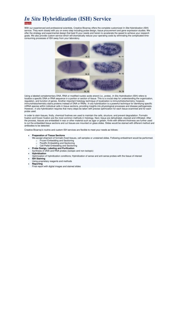
Endothelial Dystrophy Association with Hyperopia. Glenn W. Thompson, M.D. Timothy P. Page, M.D. Authors have no financial interest. Purpose. Compare the incidence of hyperopia, and refractive error of patients with and without guttata and Fuchs’ dystrophy

E N D
Endothelial Dystrophy Association with Hyperopia Glenn W. Thompson, M.D. Timothy P. Page, M.D. Authors have no financial interest
Purpose • Compare the incidence of hyperopia, and refractive error of patients with and without guttata and Fuchs’ dystrophy • Pitts and Jay first described the association between Fuchs’ dystrophy and hyperopia in 19901 • Their Fuchs’ patients had MRx of +2.48 compared to -.31 for controls, which was statistically significant • Fuchs’ patients also had shorter axial lengths and shallower anterior chambers 1. Pitts JF, Jay JL. The association of Fuchs’ corneal endothelial dystrophy with axial hypermetropia, shallow anterior chamber, and angle closure glaucoma. Br J Ophthalmol 1990:74:601-604.
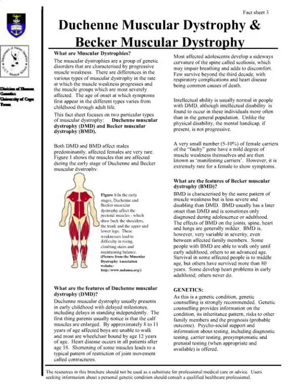
Purpose Continued • We wanted to specifically look at hyperopia related to the severity of Fuchs’ dystrophy • Stage I: cornea guttata without stromal edema • Stage II: cornea guttata with stromal edema • Stage III: stromal edema with epithelial edema and bullae, which may lead to subepithelial fibrosis
Methods • Study Population • Cases: 130 patients with 248 eyes • Controls: 127 patients with 241 eyes • Outcome Measures • Age • Gender • Family history of corneal dystrophy • Refraction including a chart review for myopic or hyperopic shifts • Visual acuity • Quantification of central corneal guttata • Presence of corneal edema
Exclusion Criteria • Previous cataract surgery with IOL was excluded unless preoperative data was available • History of refractive surgery including RK, PRK, and LASIK • Aphakic patients • Significant opacification of the ocular media preventing determination of refractive error
Demographic Data** p-value of Wilcoxon Two-Sample Test using the t Approximation
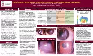
Results • A statistically significant greater proportion of study group eyes had a hyperopic spherical equivalent than the control group eyes (p-value<0.0001). • Mean spherical equivalent • Cases: +0.60 SD ± 2.65 • Controls: -0.52 SD ± 2.44 • p-value < 0.001
Refractive Results* p-value of Two-tailed Fisher’s Exact Test** p-value of Wilcoxon Two-Sample Test using the t Approximation
Fuchs’ Dystrophy Severity *p-value of Two-tailed Fisher’s Exact Test <0.0001
Conclusions • There is a significant association between the incidence of hyperopia and both the presence and degree of Fuchs’ dystrophy • This is a large study of a primarily Caucasian suburban population • Limitations of this study include the unmatched case and control age and visual acuity