Download
1 / 1
10 likes | 266 Views
In Vivo 2-Photon imaging in Synapses of Mobile Mice D. Leinweber, M. Reagan, J. Seaton, J. Sekhon Advisor: Dr. Mitch Tyler Client: Dr. Giulio Tononi and Dr. Ugo Faraguna. Final Prototype. Abstract. Dr. Tononi’s Hypothesis.

E N D
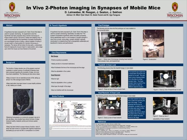
In Vivo 2-Photon imaging in Synapses of Mobile Mice D. Leinweber, M. Reagan, J. Seaton, J. Sekhon Advisor: Dr. Mitch Tyler Client: Dr. Giulio Tononi and Dr. Ugo Faraguna Final Prototype Abstract Dr. Tononi’s Hypothesis The following pictures show the final prototype as it was installed on the microscope table: A hypothesis has been proposed by Dr. Giulio Tononi that sleep is used for synaptic downsizing. Specifically, he states that “The synaptic homeostasis hypothesis claims that plastic processes during wakefulness result in a net increase in synaptic strength in many brain circuits; during sleep, synaptic strength is globally downscaled to a baseline level that is energetically sustainable and beneficial for memory and performance”. A hypothesis has been proposed by Dr. Giulio Tononi that sleep is used for synaptic downsizing. An experiment to test this hypothesis has been proposed using 2-photon microscopy to image synaptic activity in the brain in awake and sleeping mice. In order to successfully test the hypothesis of synaptic downscaling as a neurological function of sleep, a device capable of holding a mouse’s head in a fixed position for 2-photon microscopy is necessary. The device will be broken into two parts—a stereotaxic frame for keeping the mouse’s head in a rigid position, and a stage upon which the mouse can have freedom of movement. Client Requirements • Ball Design • Finish as quickly as possible • Ideally provide no movement restrictions • Must fit between the lens of the microscope and the stage • Must be adjustable in the z-plane • Head Restraint • Must be rigid • Must be adjustable in the x,y-planes • Must span the length of the stage • Must not interfere with the microscope Figure 1: Global view of microscope including head restraint, linear actuator track, and treadmill ball. Figure 2: Construction! Background Treadmill: A Closer Look The following pictures show the treadmill design in more detail: • The function of sleep remains one of the greatest unsolved mysteries in modern science. A multitude of theories have been proposed regarding the necessity of sleep, but none have been established. The following are facts about sleep: • Sleep is known to be an essential process of life, taking up almost 1/3 of the average human’s lifetime. • Sleep deprivation has been known to cause health problems in rats, leading up to death. • Retaining homeostasis is a commonly accepted view as to why we sleep, since studies have shown that lack of sleep hampers healthy metabolic activity and immune system response. • Sleep has been linked to proper memory function—lack of sleep has been shown to correlate with cognitive impairment, decreasing by as much as 38% in comparison to a control. The treadmill was made from a replaceable Styrofoam ball placed on an axel. The axel spins using a bearing bolted on to the side of a linear actuator track. The linear actuator track is used for fine adjustments to allow for different objective lengths. Figure 3: Close up view of linear actuator track. Figure 4: Close up view of treadmill ball on axel. Head Restraint: A Closer Look The following pictures show the Head Restraint design in more detail: Similar Products The head restraint was made from a steel bar spanning the gap in the stage. It was essential that the bar be adjustable, so plastic slots on the sides were used to allow for proper adjustability. Plexiglass was used to attach the plastic slots to the bar as well as to attach the entire device to the stage. Figure 6: Close up view of adjustability of head restraint. Figure 5: Close up view of head restraint. References • Dr. Ugo Faraguna, Dr. Giulio Tononi, Dr. Mitch Tyler • http://www.sciencemag.org/sciext/vis2005/show/images/slide1_large.jpg • Turner, T. H., Drummond, S. P. A., Salamat, J. S., & Brown, G. G. (2007). “Effects of 42 • hr sleep deprivation on component processes of verbal working memory.” • Neuropsychology, 21, 787-795 • Zager, A., Andersen, M. L., Ruiz, F. S., Antunes, I. B., & Tufik, S. (2007). “Effects of • acute and chronic sleep loss on immune modulation of rats.” Regulatory, • Integrative and Comparative Physiology, 293, R504-R509 • Tononi, Giulio. (2005) “Research Overview—The Center for Sleep and Consciousness.”