Download
1 / 32
320 likes | 381 Views
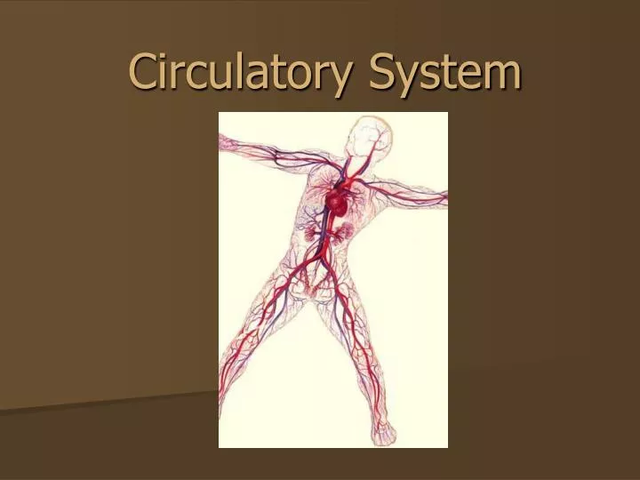
Circulatory System. The Circulatory System. Circulatory system is made up of blood, the heart, and blood vessels. Section 37.2 Summary – pages 975-984. Your Blood: Fluid Transport. Your blood is a tissue composed of fluid, cells, and fragments of cells. Components. Characteristics.

E N D
The Circulatory System Circulatory system is made up of blood, the heart, and blood vessels.
Section 37.2 Summary – pages 975-984 Your Blood: Fluid Transport • Your blood is a tissue composed of fluid, cells, and fragments of cells. Components Characteristics Transport oxygen and some carbon dioxide; lack a nucleus; contain hemoglobin Red blood cells Large; several different types; all contain nuclei; defend the body against disease White blood cells Cell fragments needed for blood clotting Platelets Liquid; contains proteins; transports red and white blood cells, platelets, nutrients, enzymes, hormones, gases, and inorganic salts Plasma • The fluid portion of blood is called plasma.
Section 37.2 Summary – pages 975-984 Your Blood: Fluid Transport • Plasma is straw colored and makes up about 55 percent of the total volume of blood. • Blood cells-both red and white-and cell fragments are suspended in plasma.
Section 37.2 Summary – pages 975-984 Red blood cells: Oxygen carriers Side view 2.0 micrometers • The round, disk-shaped cells in blood are red blood cells. Top view 7.5 micrometers • Red blood cells carry oxygen to body cells.
Section 37.2 Summary – pages 975-984 Red blood cells: Oxygen carriers • They make up 44 percent of the total volume of your blood, and are produced in the red bone marrow of your ribs, humerus, femur sternum, and other long bones. • Red blood cells remain active in the bloodstream for about 120 days, then they break down and are removed as waste.
Section 37.2 Summary – pages 975-984 Oxygen in the blood • Red blood cells are equipped with an iron-containing protein molecule called hemoglobin.
Section 37.2 Summary – pages 975-984 Oxygen in the blood • Oxygen becomes loosely bound to the hemoglobin in blood cells that have entered the lungs. • These oxygenated blood cells carry oxygen from the lungs to the body’s cells. • As blood passes through body tissues with low oxygen concentrations, oxygen is released from the hemoglobin and diffuses into the tissues.
Section 37.2 Summary – pages 975-984 Carbon dioxide in the blood • Once biological work has been done in a cell, wastes in the form of carbon dioxide diffuse into the blood and are carried in the bloodstream to the lungs.
Section 37.2 Summary – pages 975-984 White blood cells: Infection fighters • White blood cells play a major role in protecting your body from foreign substances and from microscopic organisms that cause disease. White Blood Cells • They make up only one percent of the total volume of your blood.
Section 37.2 Summary – pages 975-984 Blood clotting • Your blood contains small cell fragments called platelets, which help blood clot after an injury. • Platelets help link together a sticky network of protein fibers called fibrin, which forms a web over the wound that traps escaping blood cells.
Section 37.2 Summary – pages 975-984 Rh factor • Another characteristic of red blood cells involves the presence or absence of an antigen called RH, or Rhesus factor. • Rh factor is an inherited characteristic. • People are Rh positive (Rh+) if they have the Rh antigen factor on their red blood cells. • They are Rh negative (Rh-) if they don’t.
Section 37.2 Summary – pages 975-984 Your Blood Vessels: Pathways of Circulation • Arteries are large, thick-walled, muscular, elastic blood vessels that carry blood away from the heart. • The blood that they carry is under great pressure. • As the heart contracts, it pushes blood through the arteries.
Section 37.2 Summary – pages 975-984 Your Blood Vessels: Pathways of Circulation • Blood surges through the arteries in pulses that correspond with the rhythm of the heartbeat. • After the arteries branch off from the heart, they divide into smaller arteries that in turn divide into even smaller vessels called arterioles.
Section 37.2 Summary – pages 975-984 Your Blood Vessels: Pathways of Circulation • Capillaries are microscopic blood vessels with walls that are only one cell thick. Carries oxygenated blood. • Blood cells travel in a single file line. • Capillaries form a dense network that reaches virtually every cell in the body.
Section 37.2 Summary – pages 975-984 Your Blood Vessels: Pathways of Circulation • Thin capillary walls enable nutrients and gases to diffuse easily between blood cells and surrounding tissue cells. • As blood leaves the tissues, the capillaries join to form slightly larger vessels called venules.
Section 37.2 Summary – pages 975-984 Your Blood Vessels: Pathways of Circulation • Veins are the large blood vessels that carry blood from the tissues back toward the heart. Carries deoxygenated blood. Vein Capillary
Pulmonary Circulation – carries blood between the heart and lungs Systemic Circulation – carries blood between the heart and body Types of Circulation • Coronary Circulation– • Supplies blood to the heart
Section 37.2 Summary – pages 975-984 Your Heart: The Vital Pump • The main function of the heart is to keep blood moving constantly throughout the body.
Section 37.2 Summary – pages 975-984 Your Heart • Your heart is about 12cm by 8cm-roughly the size of your fist. • It lies in your chest cavity, just behind the breastbone and between the lungs, and is essentially a large muscle completely under involuntary control.
Superior Vena Cava OXYGEN-POOR BLOOD FLOW -Brings blood from the upper body & empties into the right atrium. To the lungs to get O2 Pulmonary Artery Pulmonary Artery To the lungs to get O2 Right Atrium Tricuspid Valve Right Ventricle Inferior Vena Cava -Brings blood from the lower body & empties into the right atrium.
OXYGEN-RICH BLOOD FLOW Left Atrium Aorta From the lungs with O2 Pulmonary Vein Pulmonary Vein From the lungs with O2 Bicuspid Valve Left Ventricle Prevents the mixing of blood Septum Aorta
Section 37.2 Summary – pages 975-984 Blood’s path through the heart • Vena Cava, Right Atrium, Right Ventricle, Pulmonary Artery, (lungs), Pulmonary Vein, Left Atrium, Left Ventricle, Aorta Superior vena cava Pulmonary artery Aorta Pulmonary vein LA RA LV RV Capillaries Inferior vena cava Left lung Right lung
Section 37.2 Summary – pages 975-984 Blood pressure • Blood pressure is the force that the blood exerts on the blood vessels. • Blood pressure rises and falls as the heart contracts and then relaxes. • Blood pressure rises sharply when the ventricles contract, pushing blood through the arteries.
Section 37.2 Summary – pages 975-984 Blood pressure • The high pressure is called systolic pressure. • Blood pressure then drops dramatically as the ventricles relax.
The Function of the Respiratory System. • Get oxygen to the cells of the body • Get rid of carbon dioxide
Parts of the Respiratory System: • PHARYNX – muscular tube in the upper throat which serves as a passageway for air and food. • LARYNX – contains your vocal chords, short passageway, sometimes called the voice box • EPIGLOTTIS – flap of tissue that covers the larynx and prevents food from going down the wrong tube. • TRACHEA - long, straight tube in the chest cavity that leads from the throat to the lungs, also called the windpipe.
Parts of the Respiratory System, cont. • BRONCHI – two small tubes that lead to the lungs, they divide into smaller tubes called BRONCHIOLES. • ALVEOLI – where the bronchioles end, clusters of air sacs. Where gases are actually exchanged. CO2->O2In healthy lungs they are elastic and can stretch, always have a small amount of air. • DIAPHRAGM – powerful muscle spanning the rib cage under the lungs, aids in respiration.
The path of oxygen and carbon dioxide through the body… Oxygen from the air diffuses into the blood vessels of the alveoli where it is used for cellular respiration inside cells. During this process, carbon dioxide diffuses into the blood where it is carried back to the lungs.