Download
1 / 24
260 likes | 480 Views
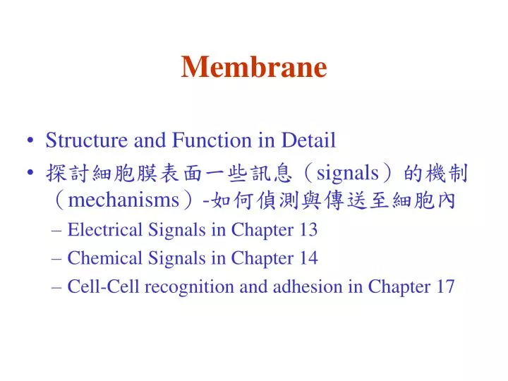
Membrane. Structure and Function in Detail 探討細胞膜表面一些訊息( signals) 的機制( mechanisms)- 如何偵測與傳送至細胞內 Electrical Signals in Chapter 13 Chemical Signals in Chapter 14 Cell-Cell recognition and adhesion in Chapter 17. Membranes: Their Structure, Function, and Chemistry. The functions of membrane

E N D
Membrane • Structure and Function in Detail • 探討細胞膜表面一些訊息(signals)的機制(mechanisms)-如何偵測與傳送至細胞內 • Electrical Signals in Chapter 13 • Chemical Signals in Chapter 14 • Cell-Cell recognition and adhesion in Chapter 17
Membranes: Their Structure, Function, and Chemistry • The functions of membrane • Models of membrane structure: An experimental perspective • Membrane lipids: The fluid part of the model • Membrane protein: The mosaic part of the model
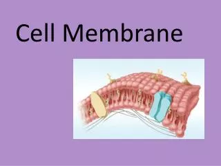
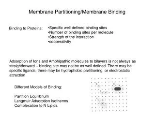
The Functions of Membranes • Membranes define boundaries and serve as permeability barrier • to define the boundaries of cell and its compartments • to serve as permeability barrier • plasma membrane and intracellular membranes • Membrane are sites of specific proteins and therefore of specific functions • Transport proteins, receptors, enzymes • Membrane proteinsregulate the transport of solutes • endocytosis and exocytosis • Membrane proteins detect and transmit electrical and chemical signals • Signal transduction • Membrane proteins mediate cell-to-cell communication • gap junctions
Membranes: Their Structure, Function, and Chemistry • The functions of membrane • Models of membrane structure: An experimental perspective • Membrane lipids: The fluid part of the model • Membrane protein: The mosaic part of the model
Models of membranes were developed long before membranes were first seen with electron microscopes in the 1950s. • In 1895, Charles Overton hypothesized that membranes are made of lipids because substances that dissolve in lipid enter cells faster than those that are insoluble. • Twenty years later, chemical analysis confirmed that membranes isolated from red blood cells are composed of lipids and proteins.
Early images from electron microscopes seemed to support the Davson-Danielli model and until the 1960s, it was considered the dominant model. • Further investigation revealed two problems. • First, not all membranes were alike, but differed in thickness, appearance when stained, and percentage of proteins to lipids. • Second, measurements showed that membrane proteins are actually not very soluble in water. • Membrane proteins are amphipathic, with hydrophobic and hydrophilic regions. • If at the surface, the hydrophobic regions would be in contact with water.
Membranes: Their Structure, Function, and Chemistry • The functions of membrane • Models of membrane structure: An experimental perspective • Membrane lipids: The fluid part of the model • Membrane protein: The mosaic part of the model
Membrane lipids: The fluid part of the model • Membranes contain several major classes of lipids • Thin-layer chromatography is an important technique for lipid analysis (TLC) • Fatty acid are essential to membrane structure and function • Membrane asymmetry: Most lipids are distributed unequally between the two monolayers • The lipid bilayer is fluid state • Membrane function properly only in the fluid state • Most organisms can regulate membrane fluidity • Lipid rafts are localized regions of membrane lipids that are involved in cell signaling
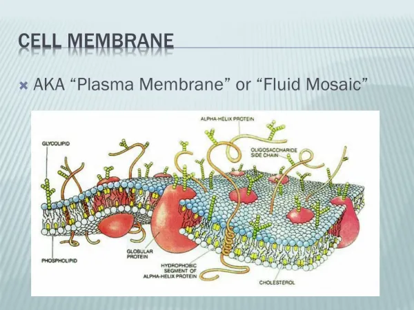
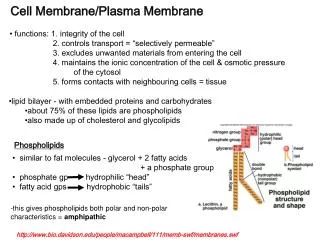
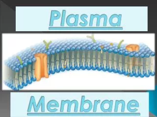
The main macromolecules in membranes are lipids and proteins, but include some carbohydrates. • The most abundant lipids are phospholipids. • Phospholipids and most other membrane constituents are amphipathic molecules. • Amphipathic molecules have both hydrophobic regions and hydrophilic regions. • The phospholipids and proteins in membranes create a unique physical environment, described by the fluid mosaic model. • A membrane is a fluid structure with proteins embedded or attached to a double layer of phospholipids.
系統組成 • 層析系統的兩個主要組成為 固定相 (stationary phase) 及 流動相 (mobile phase),二者各有不同的極性或非極性強度;樣本分子因其自身極性的強弱,與此二相之親和力不同。 與固定相親和力大者,易留滯原地; 與流動相親和力大者,易隨流動相移動,因而達成分離的目的。
Membrane Function Properly Only in the Fluid State • The effects of fatty acid composition on membrane fluidity • The effects of sterol on membrane fluidity
Most Organisms Can Regulate Membrane Fluidity • Change the lipid composition • Important for poikilotherms (變溫) : bacteria, fungi, protozoa, algae , plants, invertebrates and cold-blooded animals • Homeoviscous adaptation • To work properly with active enzymes and appropriate permeability, membrane must be fluid, about as fluid as salad oil.
Cells can alter the lipid composition of membranes to compensate for changes in fluidity caused by changing temperatures. • This allows these organisms to prevent their membranes from solidifying during winter. • For example, cold-adapted organisms, • Micrococcus (bacteria) : increase in the proportion of 16-carbon versus 18-carbon fatty acids in plasma membrane • Winter wheat(小麥): increase the percentage of unsaturated phospholipids in the autumn. • Amphibians and reptiles: increase the proportion of unsaturated fatty acid in their membranes.
Lipid Rafts(脂筏) 除了含有豐富的膽固醇及神經鞘脂類(sphingolipid) ,還包含許多細胞受體蛋白,受體蛋白可以經由與傳遞訊息的蛋白結合而活化,將細胞外的訊息傳遞到細胞內。所以脂筏被認為與傳導細胞信號通路的激活有關。
THP-1單核球細胞lipid rafts的蛋白質體學研究-徐園堤(吳烘老師) Lipid rafts 是細胞膜上的特化結構,其特性為富含cholesterol,sphingolipids與glycophosphatidylinositol-anchored proteins。在之前的研究指出,lipid rafts在物質運輸與訊息傳遞中扮演重要的角色。Lipid rafts蛋白質體上的研究大部分是利用非膠體的方法,例如使用液相層析與質譜儀。相形之下,二維電泳的方法則較少被利用來分析lipid rafts蛋白質體,儘管它能夠在ㄧ片膠上分離上千個蛋白質。本論文的研究目標主要是爲了發展一個基於二維電泳的高解析度的分離策略及結合串聯式質譜儀來建立lipid rafts的蛋白質圖譜。爲了利用二維電泳來分離lipid rafts的成分,我們以THP-1細胞為實驗模型,首先利用density gradient將lipid rafts分離出來,並使用多種以CHAPS為基礎的detergent組合來試驗二維電泳分離的效果。經蛋白質圖譜解析度與蛋白質點的數目分析結果顯示發現使用結合ASB-14與CHAPS來分離lipid rafts的效果較好。在二維電泳膠片上已利用串聯式質譜儀鑑定出17個蛋白質點,包括actin-gamma1、alpha-tubulin、G-beta1、RhoA、Rab、heat shock cognate protein、P58、GRP78以及intracellular chloride channels。由於lipid rafts與訊息傳遞有關,所以我們利用已建立的二維電泳方法來探討fractalkine處理的THP-1細胞在lipid rafts成分的變化。在lipid rafts二維電泳圖譜的影像分析顯示細胞經fractalkine刺激後導致12個蛋白質的表現出現差異。其中有6個蛋白質點的表現增加與6個蛋白質點的表現被抑制。利用二維電泳與質譜儀技術,本論文已成功地建立一個高解析度的lipid rafts蛋白質體分析平台。以蛋白質體學為基礎的策略不僅可以探索許多未知的lipid rafts蛋白質身份並且也進一步提供與lipid rafts相關的訊息傳遞路徑的了解。
Membranes: Their Structure, Function, and Chemistry • The functions of membrane • Models of membrane structure: An experimental perspective • Membrane lipids: The fluid part of the model • Membrane protein: The mosaic part of the model
Membrane Protein: The “Mosaic” Part of the Model • The membrane consists of mosaic of proteins: Evidence from freeze-fracture microscopy • Membranes contain integral, peripheral and lipid-anchored proteins • Proteins can be separated by SDS-polyacrylamide gelelectrophoresis • Molecular biology has contributed greatly to our understanding of membrane proteins • Membrane protein have a variety of functions • Membrane proteins are oriented asymmetrically across the lipid bilayer • Many membrane proteins are glycosylated • Membrane proteins vary in their mobility
Membranes contain integral, peripheral and lipid-anchored proteins • Integral membrane proteins (transmembrane protein) • Hydrophobic regions are embedded within the membrane interior. • Peripheral membrane proteins • Too hydrophilic to penetrate into the membrane but are attached to the membrane by electrostatic and hydrogen bonds to adjacent membrane. • Lipid-anchored proteins • are hydrophilic and do not penetrate into the membrane ; they are covalently bound to lipid molecules (saturated fatty acid)
Peripheral Membrane Proteins • Bind with hydrophilic portions of integral proteins • through weak electrostatic forces and hydrogen bonds with the hydrophilic portions of integral protein or with the polar head groups of membrane lipids • More readily removed from membranes than integral proteins • Extracted by changing the pH and ionic strength. • Bound to the inner surface of the plasma membrane • Form a skeleton meshwork to support the plasma membrane and maintain the shape of the erythrocyte
Lipid-Anchored Proteins • Fatty Acid-anchored Membrane Proteins (Prenylated Membrane Protein)- • Bound to inner surface • Synthesized in the cytosol • GPI-anchored Membranes (glycosylphosphatidylinositol) • synthesized in ER, transmembrane protein • Released from Phospholipase C • Attached to external surface of plasma membrane
Membrane carbohydrates are important for cell-cell recognition • The membrane plays the key role in cell-cell recognition. • Cell-cell recognition is the ability of a cell to distinguish one type of neighboring cell from another. • This attribute is important in cell sorting and organization as tissues and organs in development. • It is also the basis for rejection of foreign cells by the immune system. • Cells recognize other cells by keying on surface molecules, often carbohydrates, on the plasma membrane.
Membrane carbohydrates are usually branched oligosaccharides with fewer than 15 sugar units. • They may be covalently bonded either to lipids, forming glycolipids, or, more commonly, to proteins, forming glycoproteins. • The oligosaccharides on the external side of the plasma membrane vary from species to species, individual to individual, and even from cell type to cell type within the same individual. • This variation marks each cell type as distinct. • The four human blood groups (A, B, AB, and O) differ in the external carbohydrates on red blood cells.
Membrane Proteins Vary in Their Mobility Membrane proteins are much more variable than lipids in their mobility. Some proteins appear to move freely within the lipid bilayer whereas others are constrained, often because they are anchored to protein complex located adjacent to one side of the membrane or the other.
動物細胞融合技術 • 病毒誘導融合 • 利用仙台病毒(Sendai virus)誘導細胞融合 • 化學誘導融合 • 利用聚乙二醇(polyethylene glycol: PEG)誘導細胞融合 • PEG分子量1000左右;濃度30%40% • 電擊誘導融合