Download

1 / 51
520 likes | 709 Views
Immunity to Chlamydiae. Xi Yang Canada Research Chair in Infection and Immunity Professor Departments of Medical Microbiology and Immunology University of Manitoba For Medical Immunology, March 2013. Objectives. Chlamydial species which cause human diseases

E N D
Immunity to Chlamydiae Xi Yang Canada Research Chair in Infection and Immunity Professor Departments of Medical Microbiology and Immunology University of Manitoba For Medical Immunology, March 2013
Objectives • Chlamydial species which cause human diseases • Major diseases caused by or associated with chlamydial infections • Immune protective mechanisms • Immune pathological mechanisms • Interaction between innate and adaptive immune responses in chlamydial infection
Chlamydia • Obligate intracellular bacteria • Gram negative • Characteristic development cycle • Elementary body (infectious form) • Reticulate body (metabolic form)
The developmental cycle of Chlamydia trachomatis • Genus: Chlamydia • Chlamydia, single • Chlamydiae, plural • Not life cycle
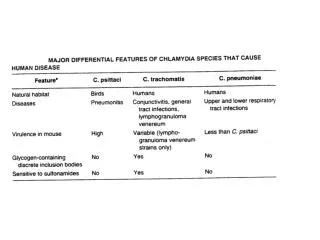
Major C. trachomatis diseases of humans Disease C. trachomatis Significance Trachomaa Serovars A, B, C Worlds leading cause of infectious preventable blindness (86 M – 3.1 M are blind or have severely impaired vision, women > risk men)c Sexually transmitted diseasesb Serovars D-K, L1-L3 92 million new cases per year globally(cervicitis, urethritis) women PID > HIV risk factor)c a One of the Worlds top seven most neglected tropical diseases b Most common bacterial caused sexually transmitted disease c WHO reports on the 2nd global scientific meeting on trachoma (2003) and global STD (2007).
Paradox of Chlamydial Host and Tissue Tropisms • MoPn vs D >99% coding ORFs conserved • D vs A >99.6% nucleotide identity Conserved and unique genes In C. trachomatis
TF (Trachomatous Inflammation-Follicular) Trachoma Normal TS (Trachomatous scarring) Mabey D. et al., Lancet, 362: 2003 TT (Trachomatous trichiasis) • Disease of increasing severity with age. • Re-infection/persistent infection drives damaging immunopathology. • Thought to be mediated by combination of ineffective protective and damaging pathologic immune responses.
The Range of Trachoma Countries with cases in 2004 Source: World Health Organization The New York Times
Fibroblasts HEV NORMAL CONJUNCTIVA 7-30 mm thick with 3 layers - Oil, aqueous, mucin defensins, PLA, Antibodies, Chemokines & cytokines Tear film Squamous epithelial cells (producing membrane tethered MUC1, 4 & 16) Goblet cells secreting mucin (MUC5AC) Stratified 3 – 7 layers thick containing goblet cells DC and IEL can traverse Basement membrane via pores Basal lamina - secreted by Epithelial cells Basement membrane Reticular lamina - secreted by fibroblasts – fibrillar collagen Lymphocyte layer Loose arrangement of lymphoid spots which contain HEV, intraepithelialLymphocytes, Langerhans cells, CD8+ T cells Lamina propria Extra cellular Matrix Random arrangement of loose packed Type I, III and IV collagen fibrils. Matrix contains Pro – IL-1b, Pro – TGF-b. Connective tissue
INFECTION & ACTIVE DISEASE Infection with Chlamydial Elementary bodies (0.3 mm) Uptake by Receptor mediated endocytosis Tear film Avoidance of phagolysosome fusion and formation of specialised inclusion (upto 1.5 mm in size) containing chlamydial reticulate bodies and Elementary bodies Thinning of stratified squamous epithelial cell layer & loss of goblet cells DC and IEL infiltrate epithelial cell layer Organisation of lymphoid spots into follicles or germinal centres which contain HEV, migratory DC, T and B Lymphocytes, plasmacytes Basement membrane HEV HEV Lymphoid Follicle Migratory DC Lymphoid follicle formation Visible to naked eye 3 –5 mm In diameter Angiogenesis B cell areas Breakdown of Extra cellular Matrix By secretion of inflammatory cytokines and MMPs. Cleavage of Pro – IL-1b Pro – TGF-b and influx of fibroblasts with collagen deposition T cell areas Fibroblasts Between follicles a diffuse mixed Infiltrate of T & B cells, Macrophage, plasma cells and neutrophils
Fibroblasts Fibroblasts Fibroblasts TRICHIASIS Disrupted tear film Tear film Infection with C. trachomatis rarely detected Squamous epithelial cell layer may be one cell thick in places Loss of normal function & increased risk of Opportunitistic secondary infection Basement membrane DC and IEL may infiltrate epithelial cell layer Poor organisation of lymphoid spots Lymphoid follicle Resolution – tissue repair and remodelling Reformation of Extra cellular Matrix Influx of fibroblasts. Non random deposition of tight bundles of types I, III & IV collagen with additional deposition of type V collagen. Fibrils are deposited on longitudinal axis anchored on posterior tarsal plate leading to loss of elasticity and tightening of tarsal plate.
NORMAL CONJUNCTIVA Goblet cells Stratified squamous epithelial cells Basement membrane Lymphocyte layer Langerhans cells CD8+ T cells IEL HEV • Vascular • Lymphocytes • Plasma cells • HEV • Follicles • Fibroblasts Lamina propria Connective tissue
ACTIVE TRACHOMA WITH LYMPHOID FOLLICLES Epithelial cell layer Deep and superficial lymphoid follicles Basement membrane HEV T cell areas B cell areas Mixed cell infiltrate
Immune responses in trachoma • Lymphoid follicle formation • Human studies in Africa: • Pathological inflammation and fibrosis associated with higher IL-4 production • Association with FoxP3+ Treg • Vaccine trial in 1960’s: killed whole organism induce partial protection (reduction of infection) but more pathology.
Challenges to trachoma vaccine development • Poor natural immunity. • Vaccine has to be superior. • Non human primates are the only animal model that mimics human infection and disease. • Protective and pathologic immune mechanisms are not well understood. • Complex host-pathogen interactions. • Infection is restricted to conjunctival epithelial cells. • Target mucosal immunity. • Extracellular infectious (EB) and intracellular replicative (RB) forms. • Represent multiple structural and secreted targets of protective immunity that could require native antigen presentation. • Recombinant subunit or acellular trachoma vaccines have been poorly efficacious in non-human primates.
Infection of the female genital tract with Chlamydia trachomatis • Genital tract infection • Pelvic inflammatory diseases (PID) • Infertility and ectopical pregnancy • Sensitive to antibiotics • Azithromycin and doxycycline. • Tetracycline and erythromycin groups
Animal models • Non-human primates • Mouse • Genital tract • Lung infection -complex of immune responses: location, species -MoPn is used most often
Immunity to genital tract infection • No vaccine • T cell immunity is most important • CD4 Th1 cells (IFNγ) are most protective • Th17?. Protective in lung model • The role of CD8 is not clear, CTL ? • Antibody is generally not protective but local IgA appears associated with protection • Antibody may play a role in prevent reinfection • Natural immunity is weak, short lasting and strain specific
protective pathologic Protective pathologic pathologic From T. Wynn
McClarty, Grant/Development of a Live-attenuated Chlamydial Vaccine Immunity To murine C. muridarum Primary Infection and Reinfection. Genital infection with C. murdiarum produces robust long-lived adaptive immunity. The immune responses that are elicited during infection resolve primary infection in approximately 4 to 5 weeks, and upon rechallenge, those adaptive immune responses result in an infection of much shorter duration (3 to 10 days), and far fewer infectious bacteria (>104 fewer) are shed. Generally speaking, adaptive immune responses elicited during infection consist of CD4+ T cells, CD8+ T cells, and antibody. CD4+ T cells are absolutely essential to bring about the resolution of primary genital infection, whereas CD8+ T cells and antibody are dispensable. In the absence of CD4+ T cells, primary infection persists. Interestingly, immunity to reinfection is governed by a more complex set of responses. First, as with primary infection, CD4+ T cells protect against reinfection and resolve infection in the absence of CD8+ T cells and/or antibody responses. However, in the context of reinfection/rechallenge, antibody is now protective and resolves secondary infection in the absence of CD4+ and/or CD8+ T cells. An indispensable element of the antibody-mediated protective immunity is the priming of the genital tract tissues by CD4+ T cells (infection-primed genital tract). Once the genital tract has been primed, CD4+ T cells are dispensable. Therefore, while the protective efficacy of antibody is dependent on CD4+ T-cell priming of the genital tract, antibody functions independently of CD4+ T cells. Adapted from Farris C M , Morrison R P Infect. Immun. 2011;79:986-996
Chlamydial antigens related to immunity and pathology • Major outer membrane proteins (MOMP) is the protective antigen which has been studied the most. Surface exposed, 60%cell wall protein. • Heat shock protein 60 appears associated with pathology • Levels of antibody to HSP60 are correlated the degree of PID. • Higher IL-10 production was detected in patients with persistent infection and PID.
Recent results of vaccination trials using Chlamydia muridarum major outer-membrane protein as antigen
Chlamydia trachomatis proteins that are recognized by human or mouse T cells
Inhibition of chlamydial growth by interferon-γ • IFNγ upregulate indoleamine 2,3 deoxygenase (IDO) expression, leading to decyclization and cellular depletion of tryptophan, an amino acid essential for chlamydial growth • Induce iNOS, nitric oxide-based killing
C. pneumoniae • High prevalence of infection (upto 60% of the population is antibody positive) • Respiratory diseases, bronchitis and pneumonia • Most are mild and asymtomatic. • Association with coronary artery and neurological diseases • CD8 cells are more important than CD4 T cells in protection
Immune cells studied in chlamydial infections • Neutrophils • NK • NKT • DC • T cell • CD4 T cell, Th1, 2, 9, 17 • CD8 T cell • γδ T cell • B cell
Interaction between innate and adaptive immune cells • γσ T cells produce IL-17 promote Th17 responses • NKT and NK cell modulate DC function for directing T cell responses • NKT promote NK cells
Experimental Strategy • Comparison between NKT-KO (V14J18 KO) and wt mice • Effect of a-GalCer administration on wt mice in following aspects: -Susceptibility to infection -CD4 and CD8 T cell responses -DC phenotype and function -DC subset function
More severe disease in NKT mice following Cpn infection D9 D15 D15 D9 D15 D9 D15 WT KO
Effect of a-Galcer on chlamydial infection and immune responses WT- GC KO- GC WT-Veh KO-Veh
Kinetics of NKT responses following Cpn infection (lung) uninfected infected NKT cytokine profile following Cpn infection (day 3)(CD1 tetramer+ CD3+)
Effect of NKT responses on T cell cytokine profile(NKT enhances IFNγ production) CD8 CD4 Cpn infection only A-GalCer + Cpn infection
Effect of NKT responses on T cell cytokine profile(NKT decreases IL-4 production) Cpn infection only A-GalCer + Cpn infection
Restoration of NKT enhance type 1 T cell responses and Cpn clearance
Conclusion (1) • NKT cells are protective in C. pneumoniae infection • NKT produces higher IFNg than IL-4, thus more NKT1-like, following Cpn infection • NKT promotes type 1 T cell response (CD4 and CD8) following C. pneumoniae infection
NKT ? NKT ? Lambrecht BN, 2001. Clin Exp Allergy
approaches Compare NKT-KO (or aGalcer treated) vs wild-type mice in: -Dendritic cell surface markers and cytokine pattern -DC function in vitro (DC:T coculture) -DC function in vivo (adoptively transfer of DC) -effect of NKT restoration in KO mice (adoptive transfer of NKT) on DC function
Altered surface phenotype and cytokine production pattern of DC in NKT-KO mice following C.pneumoniae infection.
Direct effect of NKT on DC: NKT Enhanced DC IL-12p70 production through CD40L/CD40 interaction and IFN (dependent on cell-cell contact)
Adoptive transfer to test DC function in vivo WT-DC KO-DC PBS
Adoptive transfer to test DC function in vivo (cytokine responses)
Adoptive transfer of NKT to NKT-KO mice increases IL-12 producing DC (CD8+) Restoration of NKT leads to increase of CD8a+ DC especially IL-12 producing CD8a+ DC
NKT selectively enhance IL-12p70 production by CD8+ DC subset and the effect is dependent on CD40-CD40L interaction and IFN(in vitro co-sulture)
NKT enhance the ability of CD8α+ DCs for polarizing type 1 CD8 and CD4 T cell response (in vitro) • A&B, co-culture DC subsets from infected wt and NKT mice with T cell from Cpn immunized mice • C&D, co-culture CD8+DC from infected wt mice with T cell from Cpn immunized mice
Adoptive transfer of CD8+ DCs from WT mice, but not those from NKT-KO mice, generated protective type-1 immunity in vivo.
Conclusion (II) • NKT can influence co-stimulating molecule expression on DCs • NKT can influence cytokine production patterns in DCs • NKT can modulate the function of DC in directing CD4 and DC8 T cell responses and host defense against infection • NKT can preferentially modulate CD8a+ DC subset
NKT plays important regulatory role in linking innate and adaptive immune response NKT