Download
1 / 53
530 likes | 692 Views
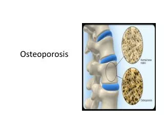
Osteoporosis Clinical Process Framework. Steven Levenson, MD, CMD. Normal and Osteoporotic Bone. The Clinical Process Framework Project. Now over a decade Started with “Green Bill” Coordinated effort between survey agency, providers, others Resulting clinical process frameworks

E N D
Osteoporosis Clinical Process Framework Steven Levenson, MD, CMD
The Clinical Process Framework Project • Now over a decade • Started with “Green Bill” • Coordinated effort between survey agency, providers, others • Resulting clinical process frameworks • Based on information in AMDA CPGs and other references and resources • A precursor to “Advancing Excellence” process frameworks
Care Process Steps • Assessment / Problem recognition • Diagnosis / Cause identification • Management / Treatment • Monitoring
OSTEOPOROSIS Clinical Process Framework • Care process step • Expectations • Rationale
Osteoporosis: Assessment / Problem Recognition • Step 1 • Did staff and physician seek and document any history of osteoporosis? • Expectations • On admission and thereafter as indicated, staff and practitioner seek and document factors associated with, or presenting risk for, osteoporosis
Step 1 Rationale • History may include • Loss of height • History of fractures (often with minimal or no trauma) • Chronic back pain due to vertebral compression fractures • Positive X-Ray finding of thinning of bone [osteopenia] • Positive bone density study (DEXA scan)
Osteoporosis: Assessment / Problem Recognition • Step 2 • Did staff identify individuals with (or risk for) osteoporosis and its complications? • Expectations • Staff and practitioner • Identify individuals with loss of bone mass and complications related to decreased bone mass • Identify and document risk factors for developing osteoporosis or for worsening of existing bone loss
Step 2 Rationale • Risk factors may be • Modifiable, for example • Inadequate calcium and vitamin D intake • Excess alcohol intake • Smoking • Medications that impair bone metabolism • Nonmodifiable, for example • Age • Female gender • Caucasian or Asian race • Small body frame
Step 2 Rationale • Various medications can increase risk of osteoporosis, for example • Anticonvulsants, proton pump inhibitors (PPIs), heparin, thyroid hormone replacement, glucocorticoids, Vitamin A
Osteoporosis In Men: Significant Risk Factors • Age (>70 years) • Low body weight (body mass index <20 to 25 kg/m2 or lower) • Weight loss (>10% compared with usual young or adult weight or weight loss in recent years) • Physical inactivity (no regular physical activity; e.g., walking, climbing stairs, housework, gardening
Osteoporosis In Men: Significant Risk Factors • Use of oral corticosteroids • Previous fragility fracture • Reference: Qaseem A, Snow V, Shekelle P, Hopkins Are, Forciea MA, Owens DK; Clinical Efficacy Assessment Subcommittee of the American College of Physicians. Screening for osteoporosis in men: a clinical practice guideline from the American College of Physicians. Ann Intern Med. 2008 May 6;148(9):680-4
Step 2 Rationale • May be benefits to addressing modifiable risk factors • Risk factors for complications include • Fall history, gait and balance disturbances, medication adverse consequences, Vitamin D deficiency
Definitions • Osteoporosis (women) • BMD that is 2.5 SD or more below the mean for women at age 30 • Osteopenia • BMD that is 1-2.5 SD below the average, for young, healthy white women. • To date, similar criteria for osteoporosis in men
Standard Deviations • Source: http://en.wikipedia.org/wiki/Standard_deviation
Hip Fracture Risks in Swedish Women • Source: www.medicographia.com
BMD Scoring • T score • Compares bone density with that of healthy young women • Z score • Compares bone density with that of other people of age, gender, and race
BMD Scanning • Also called dual-energy x-ray absorptiometry (DXA) or bone densitometry • An enhanced form of x-ray technology used to measure bone loss • Current standard for measuring bone mineral density (BMD)
BMD Scanning • DXA most often done on lower spine and hips • CT scan with special software can also be used
FRAX • Computer-based screening tool that predicts the risk of developing osteoporosis • Scoring system utilizing BMD results • Developed by World Health Organization, WHO • Can help identify individuals who should have additional testing and treatment, also depending on prognosis
Osteoporosis: Assessment / Problem Recognition • Step 3 • Did staff and practitioner identify complications of osteoporosis? • Expectations • Staff and practitioner collaborate to identify complications • Examples: impaired mobility, pain at fracture sites, deformities, deconditioning, neurological complications, psychological issues • May include in care plan document
DIAGNOSIS / CAUSE IDENTIFICATION • Step 4 • Did practitioner and staff seek causes of osteoporosis or indicate why causes could not or should not be sought?
DIAGNOSIS / CAUSE IDENTIFICATION • Expectations • Identify individuals who may benefit from additional workup • Identify any additional diagnostic workup indicated to help define presence, severity, and/or causes of decreased bone mass • Collaborate to document rationale for not screening or attempting to confirm suspected diagnosis of bone mass loss
Step 4 Rationale: Common Causes • Some medications (e.g., Dilantin, steroids) • Hyperthyroidism • Hyperparathyroidism • Chronic renal failure • Malabsorption syndromes • Multiple myeloma • Vitamin D deficiency
Step 4 Rationale: Possible Testing • Additional screening or diagnostic testing may not be needed if clinical evidence has already suggested or confirmed condition • For example, positive X-Ray showing bone thinning, a high score on a risk assessment tool, or history of vertebral compression fractures
Step 4 Rationale: Possible Testing • In absence of existing confirmation of diagnosis, presence of more advanced bone loss or significant complications may warrant screening or diagnostic testing • In absence of contraindications (e.g., terminal condition or advanced medical illness
Step 4 Rationale: Possible Testing • Depending on the situation, additional tests may include • pDEXA scan for bone density screening • Serum calcium and Vitamin D levels • TSH (hyperthyroidism) • Renal function tests (chronic renal failure)
Step 5 • Did facility identify and initiate appropriate general and specific interventions? • Expectations • Staff and practitioner institute relevant general and cause-specific interventions, or provide clinically pertinent reason for not doing so
Step 5 Rationale • Some individuals may benefit from risk reduction and cause management • Generic and cause-specific • Generic: those applicable to all at-risk individuals
Generic Interventions • Calcium (total 1200-1500 mg/day from all sources) • Vitamin D (total 800-1000 IU/day from all sources) supplementation • These may reduce additional bone loss but will not significantly improve existing bone loss
Generic Interventions • Exercise—especially weight bearing activity—may reduce bone loss • Fall prevention strategies may help reduce falls and subsequent fall-related complications of decreased bone mass
Vitamin D • Vitamin D appears to reduce fall risk • In addition to effects on bone density • Serum Vitamin D levels should be at least 24 ng/ml to reduce fall risk • Effect occurs after short duration of use • Toxicity is possible although rare • Watch for hypercalcemia • May bring out hyperparathyroidism
Step 6 • Did staff and practitioner consider possible individuals for whom additional treatment may be indicated? • Expectations • Practitioner and staff identify individuals who can benefit from additional treatments
Step 6 Rationale • Several options for medications to try to reverse bone loss • Bisphosphonates • Calcitonin • Parathyroid hormone • Hormone replacement therapy or estrogen receptor modulators • Osteoclast inhibitors • All medications for osteoporosis treatment should be prescribed and given consistent with manufacturers’ specifications and pertinent warnings related to use • Including adverse consequences and drug interactions
Step 6 Rationale • Some individuals may not be able to tolerate side effects or comply with manufacturer’s specifications for taking these medications • Do vertebroplasty and kyphoplasty help to stabilize vertebral compression fractures? • NEJM 2009; 361:557-568 - May be no more beneficial than medical pain management
Step 7 • Did staff and practitioner address complications and related risk factors? • Expectations • Staff institute relevant fall prevention strategies • Staff and practitioner identify and address symptoms such as pain related to osteoporosis or its complications
Step 7 • Expectations • Staff and practitioner evaluate patient’s current medication regimen and address medications that • Are identified or suspected as affecting bone density • May predispose to complications from osteoporosis; e.g., increase fall risk and thereby may increase risk of fracture
Step 7 Rationale • Measures to try to prevent falls and related injury may prevent injury-related complications due to osteoporosis • No interventions can prevent all falls • Sometimes necessary to focus on trying to minimize severity of complications, to extent possible
Step 8 • Did practitioner and staff follow up on individuals with osteoporosis? • Expectations • Practitioner and staff monitor progress of the condition and the individual’s response to any interventions • Based on criteria that are relevant to the individual resident
Step 8 Rationale • Sometimes difficult to identify specific long-term benefits of osteoporosis treatment in individuals • Examples of monitoring may include—as clinically appropriate—functional capacity, degree of pain, and progression, stabilization, or reduction of bone mass loss
Step 9 • Did staff and physician consider justification for continuing current approaches? • Expectations • Staff and practitioner review information that can help identify the rationale for continuing treatment
Step 9 Rationale • Various circumstances may affect decisions about continuing or modifying treatments • Prognosis • Responsiveness to treatment • Possibility for changing to a less obtrusive or lower-risk intervention • Resident satisfaction with the benefits of—or concern about complications related to—treatment
Step 9 Rationale • Reduced compliance with osteoporosis medications is common • Mostly due to adverse consequences