Download
1 / 23
230 likes | 354 Views
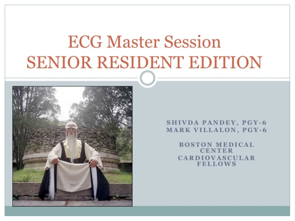
ECG Master Session SENIOR RESIDENT EDITION. Shivda Pandey , PGY-6 Mark Villalon , PGY-6 Boston Medical Center Cardiovascular fellows. What is your ECG diagnosis?. PCP Clinic Visit 65 year old male with PMH hypertension and active smoking is in your clinic for an initial evaluation.

E N D
ECG Master SessionSENIOR RESIDENT EDITION ShivdaPandey, PGY-6 Mark Villalon, PGY-6 Boston Medical Center Cardiovascular fellows
What is your ECG diagnosis? • PCP Clinic Visit • 65 year old male with PMH hypertension and active smoking is in your clinic for an initial evaluation. • He has no complaints and feels well. • Routine ECG is performed in light of his cardiac risk factors and reveals the following:
What is your diagnosis? • A. I know the diagnosis and can teach this concept to my 3rd year med student • B. I’m pretty sure about this diagnosis • C. I’m not very sure about this diagnosis • D. What does the automated read say?
Diagnosis Step 1: Normal sinus rhythm ** Always start with the rhythm**
“NSR is not “There’s a P before every QRS” • “There’s a P before every QRS” • This does not define NSR • Can be seen in flutter, a-tach etc • Sinus rhythm = Upright P’s in: • Lead I, II and aVF • Right to left activation
Diagnosis • NSR • Wow, is that QRS wide or something?
Sinus rhythm and the wide QRS 100 msec 120 msec • Normal QRS width • IVCD • “Incomplete RBBB” • “Incomplete LBBB” • LBBB • RBBB • IVCD
South Shore Plaza Fast Slow
Left Bundle Branch Block • QRS > 120 msec • V5-V6: Broad R wave • I + aVL: Absence of Q wave
RBBB: Left ventricle contracts, then right ventricle contracts • QRS > 120 msec • V1-V2: RSR’ • Lateral leads: Deep terminal S wave
A. IVCD • B. RBBB • C. LBBB
A. IVCD • B. RBBB • C. LBBB
A. IVCD • B. RBBB • C. LBBB
“When I go fast, I go wide” • Rate-related aberrancy • Usually RBBB, but can be LBBB • Refractoriness • Clinical significance: At faster rates, need to differentiate VT vs SVT with aberrancy
PA catheter insertion Pt with LBBB Complete heart block. Hopefully there’s an escape rhythm. Watch the monitor during insertion.
55M with PMH DM2 and smoking p/w 1hr of “crushing” chest pain. ECG from last week with NSR and normal QRS width. Dx? Mx? • A. New LBBB. Wait for the enzymes. • B. New LBBB. Admit to Obs unit. • C. New LBBB. Call cards fellow to activate the cath lab STAT.
55M with PMH DM2 and smoking p/w 1hr of “crushing” chest pain. ECG from last week with NSR and normal QRS width. Dx? Mx? • A. Old LBBB. Wait for the enzymes. • B. Old LBBB. Admit to Obs unit. • C. Old LBBB + acute MI. Call cards fellow STAT. • D. This is a trick question.
Discordant:QRS deflection is opposite of T wave deflection Concordant:QRS deflection is the same of T wave deflection Normal in LBBB and paced rhythm
How to diagnose an acute MI in pt with LBBB (or paced rhythm) • ST elevation ≥1 mm in a lead with upward (concordant) QRS complex - 5 points • ST elevation ≥5 mm in a lead with downward (discordant) QRS complex - 2 points • ST depression ≥1 mm in lead V1, V2, or V3 - 3 points • ≥3 points = 90% specificity of STEMI (sensitivity of 36%)