Download
1 / 42
800 likes | 2.29k Views
Approach to Chest Pain in the ED. Bryn Mumma BBB 490 October 12, 2006. Outline. Introduction History and physical exam Differential diagnosis and testing Life threatening causes Other common but not life threatening causes. Introduction.

E N D
Approach to Chest Pain in the ED Bryn Mumma BBB 490 October 12, 2006
Outline • Introduction • History and physical exam • Differential diagnosis and testing • Life threatening causes • Other common but not life threatening causes
Introduction • Roughly 5-6 million patients present with chest pain each year to EDs • The causes of chest pain range from benign to life threatening • The diagnosis remains a challenge • Difficulty lies in identifying the life-threatening without wasting resources
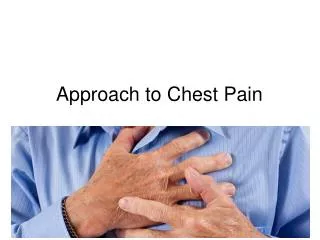
What is chest pain? • Pain in the anterior thorax, from xiphoid to suprasternal notch and between the right and left midaxillary lines. • Pain can be referred so adjacent areas are included • Character is variable – tightness, pressure, stabbing, aching, burning, etc.
Differential Diagnosis: What could it be? • Cardiac: • Myocardial ischemia • Acute myocardial infarction - Unstable angina - Stable angina • Arrhythmia • Aortic Dissection • Myocarditis/Pericarditis • Pulmonary: • Pleuritis • Pneumonia • Pulmonary embolus • Pneumothorax • Chest Wall • Cervical disc disease • Costochondritis • Herpes Zoster • Neuropathic pain • Rib fracture • Arthritis • Psychiatric • Affective disorder • Anxiety (panic attack) • Somatiform disorders • Gastrointestinal: • Esophagitis • Esophageal spasm • GERD • Esophageal rupture • Pancreatitis • Peptic Ulcer • Cholangitis • Cholecystitis • Choledocholithiasis
What information is available? • History • Physical Exam • Laboratory Tests • Imaging Tests • If at any time you are concerned of a life threatening cause of chest pain, the proper treatment should be initiated • Low risk interventions have a lower threshold
Demographics: Age, sex Chest Pain: Onset, Duration Exacerbating and Relieving factors Exercise, position Character Location Radiation Previous chest pain episodes Associated symptoms Cardiac risk factors and clotting risk factors Past medical history Previous testing Chest pain history Gives clues to cause of chest pain
Vital signs Pulse Temp Blood Pressure Respiratory Rate Oxygen Saturation Overall patient appearance Neck Veins (JVD) Cardiac auscultation Murmur, extra heart sounds Lung Auscultation Infiltrates, lung volumes, effusion, wheezing Leg swelling Chest wall or abdominal tenderness Physical Exam
Myocardial Ischemia Markers of cell injury: creatine kinase, troponin, and creatine kinase-MB Heart Failure: B-type natriuretic peptide (BNP) Pulmonary embolism D-dimer General Tests: Panel 7 Creatine Electrolytes Complete Blood Count (CBC) Anemia, elevated WBC Arterial blood gas (ABG) Ability to oxygenate Acid-base status Laboratory Tests
Imaging Tests • Electrocardiogram (ECG) • Chest x-ray • Chest CT with or without contrast • PE protocol • Dissection CT angiogram • Coronary CT angiogram • Radionuclide Perfusion Stress Test • Exercise, persantine, dobutamine • Coronary catheterization • Magnetic resonance imaging/angiography (MRI/MRA) • Echocardiography
Life-Threatening Causes • Pulmonary embolus • Tension pneumothorax • Pericarditis/cardiac tamponade • Esophageal rupture • Aortic dissection • Acute myocardial infarction
Pulmonary Embolism • Clot in the arteries leading to the lungs • Usually forms in the venous system in legs or pelvis • Approximately 500,000 patients are diagnosed with PE annually in the US, resulting in 200,000 deaths • Estimated that half of all patients with PE remain undiagnosed • Without treatment, 30% mortality rate; with proper treatment, mortality decreases to 2-8%
Pulmonary Embolism • History: Pleuritic chest pain (pain is worse when taking a deep breath), sudden onset, difficulty breathing, history of stasis, past clots, or leg swelling/pain • Exam: wheezing in the lung, rapid heart rate, low blood pressure, usually normal oxygen saturation, leg swelling (unilateral often) • Test: D-dimer, V/Q scan, chest CT • Treatment: anti-coagulation (“blood thinners”); consider thrombolytics (“clot-busters”) or surgical removable if severe
Pulmonary Embolism www.meddean.luc.edu
Tension Pneumothorax • Occurs when air can get into chest but can’t get out • Collapses lung and puts pressure on vessels/heart leading rapidly to dangerously low blood pressure • Clinical Diagnosis: sudden onset of shortness of breath, low blood pressure, and rapid heart rate; absent breath sounds over affected hemithorax; seen in young and old • Treatment: immediate needle thoracostomy to relieve pressure followed by chest tube
Tension Pneumothorax Normal Tension Pneumothorax www.scientific-com.com www.ctsnet.org
Pericarditis with tamponade • Pericarditis is an infection of the tissues surrounding the heart • Inflammation causes build-up of fluid in the closed space around the heart • History: hours to days of sharp chest pain, often positional (better when leaning forward), shortness of breath • Exam: rapid heart rate, low blood pressure, friction rub • Tests: Diffuse ECG ST segment elevation, chest x-ray, echocardiography, chest CT • Treatment: treat underlying cause, NSAIDS, drain fluid with pericardiocentesis
Esophageal rupture • Tear through the wall of the esophagus, allowing GI contents to leak into the mediastinum; usually occurs after significant vomiting or caustic ingestion • Older individual with known gastrointestinal problems. • History: Often recent violent emesis, foreign body, caustic ingestion, blunt trauma, alcoholism, esophageal disease; acute onset of localized pain • Exam: subcutaneous air (air in the soft tissue beneath the skin), decreased lung sounds • Tests: Chest x-ray, contrast esophagram, chest CT • Treatment: immediate antibiotics and surgery • 90% mortality if not treated within 24 hours
Aortic Dissection • 1 per 100,000 population with a mortality rate exceeding 90% if misdiagnosed • Large arteries have three layers • If a tear occurs in the inner vessel wall, blood can track between the layers • Artery can rupture and dissection can progress • Decreased perfusion and massive bleeding • Location determines severity
Aortic Dissection • History: Ripping/tearing chest/back pain radiating to the shoulder blade, may migrate, middle aged, high blood pressure, arterial disease • Physical: signs of blood loss (low BP, rapid heart rate), high blood pressure, ischemia, new murmur • Test: looking for markers, chest x-ray, and CT angiogram • Treatment: Medical management or surgery, depending on location and severity
Aortic Dissection MRI CT Angiogram dcmrc.mc.duke.edu
Acute Coronary Syndrome - > 500,000 deaths a year a attributed to coronary artery disease • Should be at the top of any chest pain differential • Among all chest pain patients >30yrs old • As high as 10% rate of acute myocardial infarction • As high as 25% rate of unstable angina
Myocardial Ischemia Myocardial necrosis • Ischemia is a continuum Myocardial Ischemia ST-Elevation MI Thrombus restricting blood flow Unstable Angina/Non-ST Elevation MI Narrowed vessel Stable Angina Asymptomatic CAD
What Types of Atherothrombotic Lesions Cause MI? Stable Unstable Lumen Endothelium Thrombus Platelets Lipid-Rich Core Thin Fibrous Cap Inflammatory Cells Thick Fibrous Cap MI = myocardial infarction.Adapted with permission from Falk E, et al. Circulation. 1995;92:657-671.
Acute myocardial ischemia • History: • Sudden sub-sternal crushing chest pain with radiation to the left arm/jaw • Worse with exercise (history of worsening) • Associated with shortness of breath, profuse sweating, and nausea/vomiting • Cardiac risk factors: high blood pressure, diabetes, high cholesterol, family history, tobacco use, and cocaine use • Past history of CAD/MI
Acute myocardial ischemia • Exam: • New murmur, heart sounds, elevated neck veins • Very limited utility • Testing • ECG Changes • Elevated cardiac markers • Positive stress test, cardiac cath, coronary CT angiogram
Acute myocardial ischemia • ECG Changes
Myocardial Ischemia Troponin I, CK-MB, myoglobin, and total CK are markers of cell injury Cell Death
Management ROMI = Rule Out MI Serial enzymes Serial ECG’s Telemetry monitoring Definitive testing? Research we do here may change this
Imaging – Stress Test • Identifies changes in perfusion using a radioactive tracer at rest and during exercise www.tmc.edu www.kelsey-seybold.com Tells you only about fixed defects. Does not provide information about location of blockage, degree of stenosis, or shape of thrombus.
Imaging – CT Coronary Angiogram • Timed administration of contrast dye to look at coronaries Tells you about the degree of stenosis; fast, cheap, and low risk, but another intervention is required if a blockage is seen
Imaging – Cardiac Catheterization • Higher risk • Patient must be admitted into the hospital • Can view degree of blockage and intervene www.guidant.com www.lvhhn.org
Myocardial Ischemia: Treatment • Prevent more clot from forming • Asprin (ASA), heparin, clopidogrel (Plavix), glycoprotein IIb/IIIa inhibitors, others • Increase oxygen delivery and decrease demand • Control blood pressure • Give supplemental oxygen • Pain control • Morphine • Give meds to dissolve the existing clot • Streptokinase, tissue plasminogen activator • Cardiac catheterization with percutaneous coronary intervention (angioplasty and stenting) • Coronary artery bypass graft (CABG) – open-heart bypass surgery
Myocardial ischemia: Treatment http://www.mayoclinic.com/health/coronary-angioplasty/MM00048
Other common causes • Psychiatric • Anxiety • Gastrointestinal • Acid reflux (GERD) • Esophagitis/gastritis • Musculoskeletal chest pain • Muscle strain • Costochondritis • Arthritis • Trauma • Pulmonary • Pneumonia • Asthma/COPD • Spontaneous pneumothorax • Tumor Can often be evaluated by history, exam, response to medication, and chest x-ray