Download
1 / 27
360 likes | 1.33k Views
HDR Brachytherapy. Nicolas G. Zouain, MD Department of Radiation Oncology Roger Maris Cancer Center. History. 1905. History. 1920’s. Dose Rate. Low dose rate (LDR) 0.4 to 2 Gy/hr Medium dose rate (MDR) 2 to 12 Gy/hr High dose rate (HDR)

E N D
HDR Brachytherapy Nicolas G. Zouain, MD Department of Radiation Oncology Roger Maris Cancer Center
History 1905
History 1920’s
Dose Rate • Low dose rate (LDR) • 0.4 to 2 Gy/hr • Medium dose rate (MDR) • 2 to 12 Gy/hr • High dose rate (HDR) • > 12 Gy/hr but usually in the region of 150 Gy/hr ~ 2.5 Gy/min
Why Brachytherapy • Delivering the high dose of radiation to the tumor • Sparing of the surrounding normal tissues • Delivered in a short period of time • Tumor repopulation • Limited to localized tumors
High Dose Inside of the Tumor 25% 100% 150%
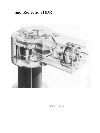
HDR Afterloading Machine Iridium-192
Lumpectomy Cavity is Created Troacar Used to Create Pathway to Cavity MammoSite Un-inflated MammoSite is Advanced into the Cavity Through the Troacar Path
Design allows the 192Ir Source to be Centrally Positioned within the Applicator The 192Ir Source is Delivered Into the MammoSite and Radiation Therapy is Delivered Per the Treatment Plan MammoSite is Inflated to Position the Tissue to Receive Radiation Therapy MammoSite