Download
1 / 38
380 likes | 530 Views
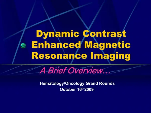
Advanced Contrast-Enhanced MRI for Stroke Risk Assessment. Bruce Wasserman, M.D. Director of Diagnostic Neurovascular Imaging Johns Hopkins Medical Institutions. Gadolinium-enhanced MR Imaging. Objectives Identifying Vulnerable Plaque (Carotid) Anatomic Features Inflammation

E N D
Advanced Contrast-Enhanced MRI for Stroke Risk Assessment Bruce Wasserman, M.D. Director of Diagnostic Neurovascular Imaging Johns Hopkins Medical Institutions
Gadolinium-enhanced MR Imaging Objectives • Identifying Vulnerable Plaque (Carotid) • Anatomic Features • Inflammation • Extend to intracranial vessels
Lumen Lipid Core Fibrous Cap Rupture Identifying Vulnerable Plaque Plaque Rupture
Lumen Lipid Core Fibrous Cap Rupture Clot Identifying Vulnerable Plaque Thin cap Large core Plaque Rupture
Black Blood MRI Lumen Plaque
Gadolinium-enhanced Black Blood MRI (CEMRI) Black Blood MRI • Enhancement of fibrous tissue improves delineation of lipid core Core Lumen Plaque - contrast MRI Post Pre
Precontrast Postcontrast H&E (Gadolinium–DPTA) Endarterectomy Specimen Lumen Calcium Fibrocellular tissue Lipid core Wasserman et al. Radiology 2002
Technical Considerations • Dedicated neck coil • 3T • 250µ in-plane resolution • 1.5T • 500µ in-plane resolution • Delayed images • 5 minutes after contrast administration • Acquire CE-MRA during injection
Contrast-Enhanced MRA (MASK images) Red-staining IPH ICA ECA Endarterectomy specimen CCA CEMRA-Mask Image MRA Qiao, Etesami, Malhotra, Astor, Virmani, Kolodgie, Trout, Wasserman. AJRN March, 2010
Intraplaque Hemorrhage ( ) IPH Detection Contrast-Enhanced MRA (MASK images) Intra- observer agreement: κ =0.94 (95% CI: 0.87-1.0) Inter-observer agreement: κ =0.91 (95% CI: 0.84-0.98) Sensitivity Specificity PPV NPV Accuracy 87% 99% 98% 91% 94% Red-staining IPH ICA ECA Endarterectomy specimen CCA CEMRA-Mask Image MRA Qiao, Etesami, Malhotra, Astor, Virmani, Kolodgie, Trout, Wasserman. AJRN March, 2010
IPH Detection Intraplaque Hemorrhage ( ) • Indicates risk for prior plaque rupture1,2 • Indicates risk for future embolic events3-6 1Chu et al. Stroke 2004;35:1079-1084 2Takaya et al. Circulation 2005;111:2768-2775 3Altaf et al. J VascSurg 2008;47:337-342 4Altaf et al. J VascSurg 2007;46:31-36 5Singh et al. Radiology 2009;252:502-508 6Takaya et al. Stroke 2006;37:818-823
61 year old male with 2 year history of left hemisphere strokes Left Carotid MRA Wasserman et al. Stroke. 2005 Nov;36(11):2504-13.
January 13, 2003 October 2003 stroke November 5, 2003 Wasserman et al. Stroke. 2005 Nov;36(11):2504-13.
November 5, 2003 T1 Precontrast T1 Postcontrast Wasserman et al. Stroke. 2005 Nov;36(11):2504-13.
T1 Precontrast T1 Postcontrast November 5, 2003 Wasserman et al. Stroke. 2005 Nov;36(11):2504-13.
Before After Sirius Red T1 Postcontrast November 5, 2003 Wasserman et al. Stroke. 2005 Nov;36(11):2504-13.
Neovascularization • Neovascularity Symptomatic, ruptured plaques1-5 • Neovascularity Intraplaque hemorrhage1 • IPH stimulates plaque progression6 1Virmani. ATVB 2005;25:2054-61 2Moreno. Circ 2004;110:2032-8 3Jeziorska. J Pathol 1999;188:189-196 4Fleiner. Circ 2004;110:2843-2850 5Russell. Br J Surg 2008;95:576-581 6Takaya. Circ 2005;111:2768-2775
Neovessels in Adventitia Postcontrast Black Blood MRI Images: Category 0: No enhancement Category 1: < 50% enhancement Category 2: ≥ 50% enhancement ICA Lumen ICA Lumen Weighted κ =0.81 (95% CI: 0.68-0.89) (Average adventitial enhancement for 5 adjacent slices) Contrast-enhancement Precontrast Postcontrast Wasserman BA. Stroke October, 2010
Association of Adventitial Enhancement with recent ipsilateralStroke/TIA Association of IPH with recent ipsilateralStroke/TIA AE Category % of cases with stroke/TIA % of cases with stroke/TIA 0 1 2 IPH absent IPH present Adventitial Enhancement P=0.001 P=0.003 (Neovascularity) 58 consecutive contrast-enhanced black blood MRI exams for carotid stenosis 2 Readers blinded to clinical information 45 males, 13 females; Mean age 73 years (49 – 89 years) 31 (53%) had recent ipsilateral stroke/TIA 32 (55%) had IPH Median stenosis = 65% Wasserman BA. Stroke October, 2010
Association of Adventitial Enhancement (AE) and IPH with Stroke/TIA IPH absent IPH present AE Category % of cases with stroke/TIA 2 Readers 0 1 2 0 1 2 Adventitial Enhancement Adventitial Enhancement Wasserman BA. Stroke October, 2010
Association of Adventitial Enhancement (AE) and IPH with Stroke/TIA Odds Ratios for Recent Stroke/TIA: IPH absent IPH present AE Category % of cases with stroke/TIA 2 Readers 0 1 2 0 1 2 Adventitial Enhancement Adventitial Enhancement Wasserman BA. Stroke October, 2010
Association of Adventitial Enhancement (AE) and IPH with Stroke/TIA Odds Ratios for Recent Stroke/TIA: Wasserman BA. Stroke October, 2010
Association of Adventitial Enhancement (AE) and IPH with Stroke/TIA Odds Ratios for Recent Stroke/TIA: Wasserman BA. Stroke October, 2010
Intracranial CE-MRI T1 FLAIR Precontrast T1 FLAIR Postcontrast T2 FRFSE Swartz RH, et al. Intracranial Arterial Wall Imaging Using High Resolution 3-Tesla Contrast-enhanced MRI. Neurology 2009;72(7):627-634.
Intracranial CE-MRI • 13 patients with intracranial atherosclerosis and recent TIA or stroke • 12 had focal eccentric wall enhancement of intracranial vessel supplying territory of acute infarct • Plaques in MCA, ACA, Basilar arteries • 10 of 12 had enhancement only in vessel supplying area of acute infarct even when multiple plaques Swartz RH, et al. Intracranial Arterial Wall Imaging Using High Resolution 3-Tesla Contrast-enhanced MRI. Neurology 2009;72(7):627-634.
Intracranial CE-MRI T1 FLAIR Precontrast T1 FLAIR Postcontrast Swartz RH, et al. Intracranial Arterial Wall Imaging Using High Resolution 3-Tesla Contrast-enhanced MRI. Neurology 2009;72(7):627-634.
May 27, 2007 64 yo male with blurred vision, dizziness and dysarthria
May 27, 2007 June 1, 2007
June 1, 2007 Sept 18, 2007 (3 months of statin use) Time
Sept 18, 2007 Time June 1, 2007 Sept 18, 2007
June 1, 2007 Sept 18, 2007 Time June 1, 2007 Sept 18, 2007
Sept 18, 2007 Feb 06, 2008 Time June 1, 2007 Sept 18, 2007 Feb 06, 2008
Sept 18, 2007 Feb 06, 2008 Feb 06, 2008 Sept 18, 2007 Feb 06, 2008 Time
Summary Contrast-enhanced Plaque MRI: • Black blood images pre- and post-contrast • Fibrous cap thickness, lipid core size, calcification • Adventitial enhancement score (i.e., neovascularity) • CEMRA during arterial phase • Mask image - IPH
Summary Contrast-enhanced Plaque MRI: Determine plaque vulnerability Identify the culprit lesion Monitor therapeutic response
Acknowledgments • Ye Qiao, PhD • MaryamEtesami, MD • Brad Astor, PhD • Matthias Stuber, PhD • Steven R. Zeiler, MD, PhD • Robert Wityk, MD • Victor Urrutia, MD • Hugh H. Trout, III, MD • Frank Kolodgie, PhD • RenuVirmani, MD