Download
1 / 39
400 likes | 628 Views
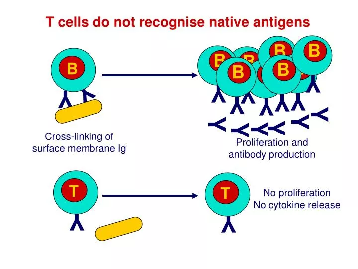
B. B. B. B. B. B. B. B. B. Y. Y. Y. Y. Y. Y. Y. Y. Y. Y. T. T. Y. Y. T cells do not recognise native antigens. Y. Y. Y. Y. Y. Y. Cross-linking of surface membrane Ig. Proliferation and antibody production. No proliferation No cytokine release. T. Y.

E N D
B B B B B B B B B Y Y Y Y Y Y Y Y Y Y T T Y Y T cells do not recognise native antigens Y Y Y Y Y Y Cross-linking of surface membrane Ig Proliferation and antibody production No proliferation No cytokine release
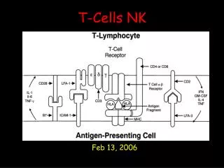
T Y Cell surface peptides of Ag presented by cells that express MHC antigens Soluble native Ag Soluble peptides of Ag Cell surface peptides of Ag Cell surface native Ag Antigens must be processed in order to be recognised by T cells ANTIGEN PROCESSING T cell response No T cell response No T cell response No T cell response No T cell response
M M M M Early evidence that antigens are catabolised Macrophages and radiolabelled Listeria monocytogenes Rapid binding to cell surface Degradation of bacteria and release of Radiolabelled protein into supernatant and cells Internalisation How is antigen catabolism linked to T cell proliferation?
Listeria-specific T cells NO T CELLS BIND T Listeria coated plastic M M NO T CELLS BIND NO T CELLS BIND NO T CELLS BIND T CELLS BIND 0mins 60mins M M The interaction of T cells with macrophages requires antigen catabolism Listeria T cell do not bind stably to antigen presenting cells unless the antigen is catabolised
Listeria-specific T cells T M M M Only metabolically active cells can process antigen Fix with paraformaldehyde or poison with sodium azide Pulse with Listeria for 60min & wash cells Add Listeria specific T cells NO T CELLS BIND Determinants recognised by T cells are generated by catabolic activity that is dependent upon the viability of macrophages Antigen presenting cells must be viable to PROCESS antigen
T M M Add Listeria specific T cells M M M Antigen presentation does not require metabolically-active cells Listeria Fix with paraformaldehyde or poison with sodium azide T CELLS BIND Antigen presenting cells do not need to be viable to PRESENT antigen
Listeria T M M M M Listeria Add Listeria specific T cells M M M M Where does antigen processing take place? T CELLS BIND Incubate with CHLOROQUINE NO T CELLS BIND Chloroquine inhibits lysosomal function (a lysosomotrophic drug) Antigen processing involves the lysosomal system
Ovalbumin specific T cell line T T T T T T T T T T T T T T T T T Native ovalbumin Digested ovalbumin Ag APC APC APC APC APC Viable Viable Fixed Fixed T cell response What form of antigen is produced by antigen processing? Catabolism reduces antigens to peptides that can be recognised by T cells
Summary of exogenous antigen processing • T cells can not recognise native antigens • Antigens must be processed for recognition by T cells • Antigens catabolism occurs inside cells • Only metabolically active cells can process antigen • Antigen presentation does not require metabolically-active cells • Antigen processing involves the lysosomal system • Catabolism reduces antigens to peptides • Because extracellular antigens are dealt with by the lysosomal system, lysosomal antigen processing is part of the EXOGENOUS antigen processing pathway
• Macrophages have well- developed lysosomal systems MF Is exogenous antigen processing sufficient? • Specialised for motility, phagocytosis and the introduction of particles to the lysosomal system Most cell types do not have lysosomal systems developed as well as macrophages BUT Viruses can infect most cell types A non-lysosomal mechanism to process antigens for presentation to T cells is required
Infectious influenza CTL assay Kill CTL CTL CTL CTL CTL No treatment Cloned anti- CTL Kill CTL CTL CTL CTL CTL CTL CTL CTL CTL + Chloroquine CTL CTL Infectious viruses raise CTL that recognise antigens that are not generated by the exogenous pathway Strong T cell response Lysosome inhibitors do not inhibit the generation of antigens recognised by most CTL Most CTL do not recognise lysosomally-derived antigens
Inactivated influenza CTL assay Weak T cell response Kill CTL Cloned anti- CTL No treatment CTL CTL CTL CTL CTL CTL CTL CTL CTL CTL CTL + Chloroquine Inactive viruses raise CTL to antigens that are generated by the exogenous pathway No Kill Lysosomal inhibitors inhibit the generation of antigens from INACTIVE virus Some CTL can recognise lysosomally-derived antigens
CTL raised with infectious virus CTL raised with non-infectious virus CTL CTL Untreated Protein synthesis inhibitor-treated Non-lysosomal processing The antigens of infectious & inactivated viruses are clearly generated by different mechanisms Infectious viruses use cellular protein synthesis machinery to replicate Inactivated viruses do not synthesise protein Protein synthesis is required for virus infected target cells to express antigens recognised by CTL
Non-lysosomal antigen processing Inactive virus raises a weak CTL response The processing of antigens from inactive viruses is sensitive to lysosomotrophic drugs ANTIGENS FROM INACTIVE VIRUSES ARE PROCESSED VIA THE EXOGENOUS PATHWAY Infectious virus raises a strong CTL response The processing of antigens from infectious viruses is NOT sensitive to lysosomotrophic drugs Most CTL recognise antigens generated via a non-lysosomal pathway Protein synthesis is required for non-lysosomal antigen processing ANTIGENS FROM INFECTIOUS VIRUSES ARE PROCESSED VIA THE ENDOGENOUS PATHWAY Do the two pathways generate the same type of T cell receptor ligand?
Influenza virus Peptides of nucleoprotein Nucleoprotein CTL CTL CTL Native antigen fails to sensitise for lysis No protein/antigen synthesis Endogenous antigen processing also generates peptides Infectious virus sensitises for lysis Protein/antigen synthesis Synthetic peptide antigens sensitise targets for lysis No protein/antigen synthesis but peptides are pre-formed
Y The site of pathogen replication or mechanism of antigen uptake determines the antigen processing pathway used EXTRACELLULAR OR ENDOSOMAL REPLICATION Y Vesicular Compartment Contiguous with extracellular fluid Exogenous processing (Streptococcal, Mycobacterial antigens) INTRACELLULAR REPLICATION Cytosolic compartment Endogenous processing (Viral antigens) Distinct mechanisms of antigen generation are used to raise T cells suited to the elimination of endogenous or exogenous pathogens
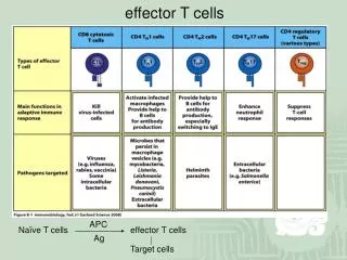
Y Antigens generated by endogenous and exogenous antigen processing activate different effector functions EXOGENOUS PATHOGENS ENDOGENOUS PATHOGENS Y Eliminated by: Antibodies and phagocyte activation by T helper cellsthat use antigens generated by EXOGENOUS PROCESSING Eliminated by: Killing of infected cells byCTL that use antigens generated by ENDOGENOUS PROCESSING
Stages of endogenous and exogenous antigen processing UPTAKE Access of native antigens and pathogens to intracellular pathways of degradation DEGRADATION Limited proteolysis of antigens to peptides ANTIGEN-MHC COMPLEX FORMATION Loading of peptides onto MHC molecules ANTIGEN PRESENTATION Transport and expression of peptide-MHC complexes on the surface of cells for recognition by T cells
Y Y Y Uptake of exogenous antigens Membrane Ig receptor mediated uptake Y Phagocytosis Complement receptor mediated phagocytosis Pinocytosis Fc receptor mediated phagocytosis Uptake mechanisms direct antigen into intracellular vesicles for exogenous antigen processing
Non-receptor -mediated uptake Receptor-mediated antigen uptake 100 75 50 25 0 10-3 10-2 10-1 Receptor-mediated uptake enhances the efficiency of the T cell response % of max. T cell response Antigen gml-1
Cell surface Uptake Endosomes Increase in acidity To lysosomes Exogenous pathway Protein antigens In endosome Cathepsin B, D and L proteases are activated by the decrease in pH Proteases produce ~24 amino acid long peptides from antigens Drugs that raise the pH of endosomes inhibit antigen processing
MHC class II maturation and invariant chain In the endoplasmic reticulum Invariant chain stabilises MHC class II by non- covalently binding to the immature MHC class II molecule and forming a nonomeric complex Need to prevent newly synthesised, unfolded self proteins from binding to immature MHC
and b chains of MHC class II molecules CLIP Invariant chain CLIP peptide A peptide of the invariant chain blocks the MHC molecule binding site. This peptide is called the CLass II associated Invariant chain Peptide (CLIP)
Cell surface Endosomes Uptake Class II associated invariant chain peptide (CLIP) (inv)3 complexes directed towards endosomes by invariant chain Cathepsin L degrades Invariant chain CLIP blocks groove in MHC molecule MHC Class II containing vesicles fuse with antigen containing vesicles
Removal of CLIP ? How can the peptide stably bind to a floppy binding site? Competition between large number of peptides
HLA-DM assists in the removal of CLIP HLA-DM HLA-DR HLA-DM: Crystallised without a peptide in the groove In space filling models the groove is very small
HLA-DM Single pocket in “groove” insufficient to accommodate a peptide HLA-DR Multiple pockets in groove sufficient to accommodate a peptide
HLA-DM HLA-DR Sequence in cytoplasmic tail retains HLA-DM in endosomes HLA-DM catalyses the removal of CLIP HLA-DM Replaces CLIP with a peptide antigen using a catalytic mechanism (i.e. efficient at sub-stoichiometric levels) Discovered using mutant cell lines that failed to present antigen HLA-DO may also play a role in regulating DM MIIC compartment
Exported to the cell surface (t1/2 = 50hr) Sent to lysosomes for degradation Surface expression of MHC class II- peptide complexes MIIC compartment sorts peptide-MHC complexes for surface expression or lysosomal degradation
Endogenous antigen processing UPTAKE Antigens/pathogens already present in cell DEGRADATION Antigens synthesised in the cytoplasm undergo limited proteolytic degradation in the cytoplasm ANTIGEN-MHC COMPLEX FORMATION Loading of peptide antigens onto MHC class I molecules is different to the loading of MHC class II molecules PRESENTATION Transport and expression of antigen-MHC complexes on the surface of cells for recognition by T cells
Degradation in the proteasome Cytoplasmic cellular proteins, including non-self proteins are degraded continuously by a multicatalytic protease of 28 subunits The components of the proteasome include MECL-1, LMP2, LMP7 These components are induced by IFN- and replace constitutive components to confer proteolytic properties. LMP2 & 7 encoded in the MHC Proteasome cleaves proteins after hydrophobic and basic amino acids and releases peptides into the cytoplasm
Newly synthesised MHC class I molecules Peptides need access to the ER in order to be loaded onto MHC class I molecules Peptide antigens produced in the cytoplasm are physically separated from newly formed MHC class I ENDOPLASMIC RETICULUM CYTOSOL
Hydrophobic transmembrane domain Lumen of ER Lumen of ER Peptide Peptide Peptide Peptide Peptide Peptide Peptide Peptide Peptide Peptide Peptide ER membrane ER membrane TAP-1 TAP-1 TAP-1 TAP-1 TAP-1 TAP-1 TAP-1 TAP-1 TAP-1 TAP-1 TAP-1 TAP-2 TAP-2 TAP-2 TAP-2 TAP-2 TAP-2 TAP-2 TAP-2 TAP-2 TAP-2 TAP-2 Cytosol Cytosol ATP-binding cassette (ABC) domain Peptide antigens from proteasome Transporters associated with antigen processing (TAP1 & 2) Transporter has preference for >8 amino acid peptides with hydrophobic C termini.
Analysis of genes in the MHC of the mutant cell line showed mutations in a pair of ABC transporter genes Normal antigen presenting cell line with stable surface MHC class I expression X √ Chemically-induced mutant antigen presenting cell line with unstable (floppy) MHC class I expressed intracellularly Discovery of the role of TAP1 & TAP2 in antigen processing Transfection of normal TAP genes into mutant APC restored stable surface MHC class I expression Mutations in TAP genes affect the supply of peptides to the ER MHC class I stability is dependent upon a supply of peptides
Peptide Peptide Peptide Peptide Peptide Peptide Peptide Peptide Peptide Peptide Peptide TAP-1 TAP-1 TAP-1 TAP-1 TAP-1 TAP-1 TAP-2 TAP-2 TAP-2 TAP-2 TAP-2 TAP-2 TAP-2 TAP-2 TAP-2 TAP-2 TAP-2 TAP-1 TAP-1 TAP-1 TAP-1 TAP-1 Endoplasmic reticulum Maturation and loading of MHC class I B2-M binds and stabilises floppy MHC Tapasin, calreticulin, TAP 1 & 2 form a complex with the floppy MHC Calnexin binds to nascent class I chain until 2-M binds Cytoplasmic peptides are loaded onto the MHC molecule and the structure becomes compact
Exported to the cell surface Sent to lysosomes for degradation Fate of MHC class I
Peptide Peptide Peptide Peptide TAP-1 TAP-1 TAP-1 TAP-2 TAP-2 TAP-2 TAP-2 TAP-2 TAP-1 TAP-1 Endoplasmic reticulum Sent to lysosomes for degradation HSV protein blocks transport of viral peptides into ER Evasion of immunity by interference with endogenous antigen processing
Normally exported to the cell surface Adenoviral protein retains MHC class I in the ER Sent to lysosomes for degradation Evasion of immunity by interference with endogenous antigen processing
Summary • T and B cells recognise antigen differently • Antigen must be catabolised before T cells can recognise it • Antigen processing generates antigenic peptides • Exogenous antigen processing takes place in lysosomes • Endogenous processing is non-lysosomal • The mechanism of antigen processing depends upon the compartment in which the pathogen replicates • Endogenous and exogenous antigen processing both involve uptake, degradation, complex formation and presentation • Exogenous antigen processing uses invariant chain and HLA-DM • Endogenous antigen processing uses proteasomes and peptide transporters in antigen processing • Pathogens can evade immunity by disrupting antigen processing