Download
1 / 34
350 likes | 551 Views
The role of the apex in differentiation growth: The development of leaves. The foliar buttress and the formation of new leaves. The foliar buttress develops in close proximity to the apex.

E N D
The role of the apex in differentiation growth: The development of leaves
The foliar buttress and the formation of new leaves The foliar buttress develops in close proximity to the apex The leaf expands rapidly, in width and in length, through division of meristematic cells called initials
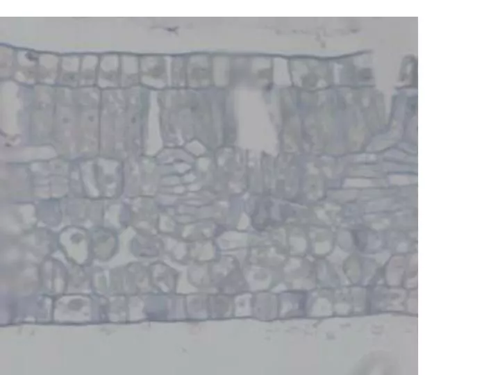
Becoming different -- differentiation Newly-formed cells are initially very similar to each other, but signs of differentiation can soon be seen. This image is a section of part of a leaf and has been sectioned parallel to the surface, so that we can see part of the developing vascular network. Veins are formed by a special subgroup of meristematic cells, called the submarginal initials.
The two groups of initials MARGINAL INITIALS:Responsible for the formation of the epidermis & hypodermis SUBMARGINAL INITIALS: Responsible for the formation of the mesophyll and by a separate route, the VASCULAR TISSUE
The influence of photosynthetic type on leaf differentiation Whether the plant is a C3, or a C4, or CAM, photosynthetic type will affect the shape, size and internal structure (development; differentiation) of the leaf. In CAM plants, water conservation is critical and spongy mesophyll is centrally-located and stores water In C3 plants, chloroplasts structure is the same in all photosynthetic tissue. In C3 dicots chloroplasts are di --> polymorphic, if mesophyll is differentiated then this forms into palisade and spongy mesophyll, else undifferentiated mesophyll. In C4 monocots, mesophyll is differentiated into Kranz and non-Kranz mesophyll. In C4 dicots, the mesophyll is also differentiated, this time into non-Kranz and Kranz mesophyll
Adaxial epidermis mesophyll between bundles is undifferentiated Bundle sheath xylem SI MI Vascular tissue procambium phloem Kranz mesophyll Abaxial epidermis Leaf differentiation in a C4 monocot
Adaxial epidermis Adaxial palisade mesophyll Bundle sheath xylem MI SI Vascular tissue procambium phloem Bundle sheath Abaxial palisade mesophyll Abaxial epidermis Leaf differentiation in a C3 dicot
MI SI Controlling development and differentiationdicotyledonous foliage leaves adaxial epidermis adaxial palisade mesophyll vascular bundles procambium bundle sheath (parenchymatous) abaxial (spongy) mesophyll abaxial epidermis
adaxial palisade mesophyll (non-Kranz) bundle sheath procambium MI SI bundle sheath abaxial palisade mesophyll (non-Kranz) Controlling development and differentiationdicotyledonous foliage leaves C4 adaxial epidermis Kranz mesophyll vascular bundles Kranz mesophyll abaxial epidermis
So, how does differentiation work? Where do cells originate? Where do tissues form?
AM Domains in apical development The apical meristem is one of the simplest-looking structures in the higher plant, yet, the processes controlling its differentiation sequencing is not yet fully understood. We recognize that changes have to be effected in the way in which neighbouring cells communicate (or stop communicating) prior to, during and after a cell division event in this structure. This topic explores the concept of domain control in higher plants, specifically in the shoot apex.
simplex monoplex duplex Shoot apical meristem type – increasing complexity Here, all subsequent cells are related to one single AM cell. Common in lower order plants. Here several AM cells are involved in production of new initials and derivatives – however, zonation becomes apparent and easier to explain. Here a number (possibly three) AM cells are involved in the formation of new initials and derivatives.
monoplex NB: ANTICLINAL means perpendicular to a surface; PERICLINAL is parallel to a surface. = plane of division monoplex Monoplex shoot apical meristems have a single top cell, often tetrahedral and produces daughter cells by lateral cell division. A relatively simple structure, where all cells have direct lineage to the apical mother cell. Separation into cortex and stele requires isolation of derivatives to allow for periclinal and anticlinal cell division d1l d2l d2r d3 d2 d1r d1
simplex simplex The simplex apical meristem has a zone of initials in an unstable sub-superficial layer. Cells may divide in the horizontal and the vertical plane. Not all cell have the same lineage. A slightly more complex structure can evolve than in monoplex systems. How does it function?
simplex Alternative division plane The working simplex zone 1
The duplex The duplex apical meristem has two layers of sub-superficial cells. These give rise to two lineage compartments – the tunica and corpus. This results in an apical meristem with two distinct cellular features (recognizable quite early on in development) and will give rise, through the to the two major cell lineages, to the cortex and the stele, and its associated tissues.
outer zone domain 2 inner zone domain 1 The duplex – a “black box” – two domains This system (common in higher plants) allows for independent cell division in the two compartments. It is initiated through closed-gating of plasmodesmata. = plasmodesma closed
2 2 1 2 1 2 1 construction…and the need for continuity .. sometimes! 1 1 1 1
symplasmic continuity tunica (CZT) peripheral tunica (CZPT) CZT = cell zone: tunica CZC = cell zone: corpus CZTP=lateral cell zone: peripheral tunica corpus CZC Three zones can be recognized within the apex: (1). the tunica, (2) the peripheral tunica zone and (3) the central corpus zone. All are in symplasmic contact. This is thus a singledomain.
(2) tunica and corpus symplasmically connected tunica (CZT) peripheral tunica (CZPT) corpus CZC symplasmic continuum here, means that all the cells are in contact and that small moleculesand signals may be transported through all cells in the developing apex, via plasmodesmata. The concept of a signal gradient can be argued.
(3) tunica in symplasmic continuity, corpus isolated tunica (CZT) = plasmodesma closed CZT = cell zone: tunica CZC = cell zone: corpus CZTP=cell zone: peripheral tunica peripheral tunica (CZTP) Here, tunica as well as peripheral tunica are symplasmically connected, but isolated from the corpus. Corpus could engage in non-synchronous cell division, to produce cells withoutthe influence of the tunica. corpus (CZC)
= plasmodesma closed (4) Tunica has role in mediating in symplasmic continuity if corpus isolated, division processes signaled tunica (CZT) peripheral tunica (CZPT) CZT = cell zone: tunica CZC = cell zone: corpus CZTP=cell zone: peripheral tunica corpus (CZC)
= plasmodesma closed (5) Zonation: When a CZPT region becomes isolated New event can occur tunica (CZT) Signal gradient isolation Signal gradient peripheral tunica (CZPT) CZT = cell zone: tunica CZC = cell zone: corpus CZTP=cell zone: peripheral tunica corpus (CZC) Here….1/23/2008
Summary:The apex, simple cells, complex arrangement, new form and function Apical meristem
epidermal and subepidermal development – step one establishing gradients
Conclusion: It is possible to apply this model to the development of a leaf as well. Clearly, Cell division can be synchronous (cell compartments in harmony) or asynchronous (dividing cell compartments isolated). Synchrony or asynchrony can thus determine the (a) type of derivative cell formed (b) the type of tissue formed and its position. So what happens in the apex is that the puzzle pieces are simply(!) put together and orchestrated during the early developmental stages…. tunica (CZT) peripheral tunica (PTZ) corpus CZC Plasmodesma are the key
An extension of and to, the regulatory pathway? Whether we deal with the apex or a developing leaf, it makes good sense to recognise that domains exist in mature tissues and that these domains are functional and operate to regulate not only the flow of information, but also, the flow of assimilates into the phloem in a source leaf.
Spheres of influence – movement of signals? This diagram shows that there is a degree of influence possible if there are overlapping domains in our system. The points of ‘overlap’ – (really domain boundaries) will possibly influence neighbouring cells under specific conditions, and at set point during the development of new cells within the duplex apical meristem. The red and blue arrows simply show two possibilities for a multidirectional signalling potential.