Download
1 / 19
190 likes | 402 Views

E N D
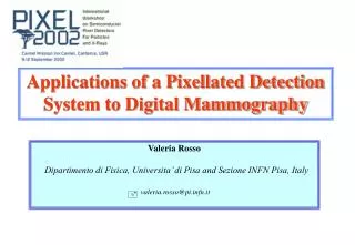
1. Physical & Psychophysical Evaluation of a New Digital Specimen Radiography System for Use in Mammography Elizabeth Krupinski, PhD
Hans Roehrig, PhD
University of Arizona
William Schempp, PhD
MedOptics Corporation
2. Rationale Specimen radiography of excised breast tissue is critical to ensure that an appropriate tissue sample has been obtained or that complete removal of the lesion has been successful.
3. Objectives To physically characterize the new high-resolution digital specimen radiography system from MedOptics Corporation.
To evaluate human observer detection performance using images acquired with the new system.
4. The Digital System Camera
1024 x 1024, 1� x 1� large area high-res CCD
2:1 fiber optic taper bonded to CCD (2�x2� FOV)
standard Lanex Regular screen coupled to large end of taper
image data digitized at 500 Kpix/sec, 12-bits
Faxitron MICRO 50 x-ray source
15-50 kVp range
1-90 sec time selection
SID = 50 cm
5. Faxitron HVL & Eeff
6. Camera X-radiation Response
7. Camera Dark Signal & Noise
8. Noise Power Spectrum 25 kVp
9. Noise Power Spectrum 30 kVp
10. Comparison NPS & Square MTF
11. MTF
12. MTF from Square Wave Response
13. Observer Performance Study 2 CDMAM contrast-detail phantoms
Commercially available large version
Small experimental version
Imaging conditions
Traditional plain film
Fuji Computed Radiography (CR)
MedOptics Digital System
25 kV & 30 kV @ High (3600 GL), medium (2400 GL) & low (1200 GL) exposures
14. Film Image Optical Densities
15. Viewing Conditions Traditional film & Fuji CR film
standard viewbox @ 1100 ft-L
extraneous light masked
Fuji CR digital & MedOptics Digital
DataRay DR110
1728 x 2304
140 ft-L
Perceptually linearized with Barten curve
16. Observers & Task 10 undergrad students in each condition
On outline of phantom indicate location of all disks visible in the phantom(s)
Window/Level & zoom available on monitor
Ambient lights turned off in all conditions
17. Large Phantom Results
18. Small Phantom Results
19. Performance Summary Increases monotonically low to high exposures
25 kV > 30 kV
MedOptics Digital System Best
Traditional plain film worst
Fuji CR monitor & film in middle
Minor differences between phantoms
20. Conclusions MedOptics Digital Biopsy System yields high detection performance
Currently mammographers image core biopsy samples with either traditional systems or stereotactic units
Traditional takes too long
Stereotactic requires patient to get out of unit to image sample
Digital avoids these problems & is fast