Download
1 / 76
760 likes | 985 Views
Veins. Collect blood from all tissues and organs and return it to the heart Are classified according to size Venules Medium-sized veins Large veins. Arterial blood pressure. Arterial blood pressure Maintains blood flow through capillary beds

E N D
Veins • Collect blood from all tissues and organs and return it to the heart • Are classified according to size • Venules • Medium-sized veins • Large veins
Arterial blood pressure • Arterial blood pressure • Maintains blood flow through capillary beds • Rises during ventricular systole and falls during ventricular diastole • Pulse is a rhythmic pressure oscillation that accompanies each heartbeat • Pulse pressure = difference between systolic and diastolic pressures Mean arterial pressure (MAP)
Figure 21.10 Pressures within the Cardiovascular System Figure 21.10
Figure 21.10 Pressures within the Cardiovascular System Figure 21.10
Capillary Exchange • Flow of water and solutes from capillaries to interstitial space • Plasma and interstitial fluid are in constant communication • Assists in the transport of lipids and tissue proteins • Accelerates the distribution of nutrients • Carries toxins and other chemical stimuli to lymphoid tissues
Processes that move fluids across capillary walls • Diffusion • Filtration • Hydrostatic pressure (CHP) • Reabsorption
Figure 21.12 Capillary Filtration Figure 21.12
Figure 21.12 Capillary Filtration Figure 21.12
Forces acting across capillary walls • Capillary hydrostatic pressure (CHP) • Blood colloid osmotic pressure (BCOP) • Interstitial fluid colloid osmotic pressure (ICOP) • Interstitial fluid hydrostatic pressure (IHP)
Filtration and reabsorption • Processes involved in filtration and reabsorption include • Net hydrostatic pressure • CHP - IHP • Net colloid osmotic pressure • BCOP - ICOP
Figure 21.13 Forces Acting across Capillary Walls Figure 21.13
Venous pressure and venous return • Assisted by two processes • Muscular compression • The respiratory pump PLAY Animation: Factors that Affect Blood Pressure
Cardiovascular Regulation • Autoregulation • Neural mechanisms • Endocrine mechanisms
Autoregulation of blood flow within tissues • Local vasodilators accelerate blood flow in response to: • Decreased tissue O2 levels or increased CO2 levels • Generation of lactic acid • Release of nitric acid • Rising K+ or H+ concentrations in interstitial fluid • Local inflammation • Elevated temperature
Figure 21.14 Homeostatic Adjustments that Compensate for a Reduction in Blood Pressure and Blood Flow Figure 21.14
Figure 21.14 Homeostatic Adjustments that Compensate for a Reduction in Blood Pressure and Blood Flow Figure 21.14
Neural Mechanisms • Adjust CO and PR to maintain vital organ blood flow • Medullary centers of regulatory activity include • Cardiac centers • Vasomotor centers control • Vasoconstriction via adrenergic release of NE • Vasodilation via direct or indirect release of NO
Reflex control of cardiovascular function • Baroreceptors reflexes monitor stretch • Atrial baroreceptors monitor blood pressure • Chemoreceptor reflexes monitor CO2, O2, or pH levels PLAY Animation: Autoregulation and Capillary Dynamics
Figure 21.15 Baroreceptor Reflexes of the Carotid and Aortic Sinuses Figure 21.15
Figure 21.16 The Chemoreceptor Reflexes Figure 21.16
Hormones and cardiovascular regulation • Antidiuretic hormone – released in response to decreased blood volume • Angiotensin II – released in response to a fall in blood pressure • Erythropoietin – released if BP falls or O2 levels are abnormally low • Natriuretic peptides – released in response to excessive right atrial stretch
Figure 21.17 The Regulation of Blood Pressure and Blood Volume Figure 21.17a
Figure 21.17 The Regulation of Blood Pressure and Blood Volume Figure 21.17b
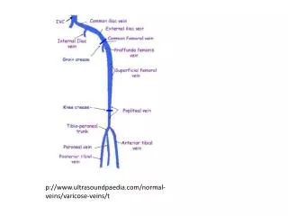
Venous Valves • Venules and medium-sized veins contain valves • Prevent backflow of blood
Figure 21.6 The Function of Valves in the Venous System Figure 21.6
Distribution of blood • Total blood volume is unevenly distributed • Venoconstriction maintains blood volume • Veins are capacitance vessels • Capacitance = relationship between blood volume and pressure PLAY Animation: Anatomy Review: Blood Vessel Structure and Function
Figure 21.7 The Distribution of Blood in the Cardiovascular System Figure 21.7
Circulatory Pressure • Circulatory pressure is divided into three components • Blood pressure (BP) • Capillary hydrostatic pressure (CHP) • Venous pressure
Figure 21.8 An Overview of Cardiovascular Physiology Figure 21.8
Resistance (R) • Resistance of the cardiovascular system opposes the movement of blood • For blood to flow, the pressure gradient must overcome total peripheral resistance • Peripheral resistance (PR) is the resistance of the arterial system
Overview of Cardiovascular Pressures • Factors involved in cardiovascular pressures include • Vessel diameter • Cross-sectional area of vessels • Blood pressure • Blood viscosity
Figure 21.9 Relationships among Vessel Diameter, Cross-sectional Area, Blood Pressure, and Blood Viscosity Figure 21.9
Exercise and the Cardiovascular System • Light exercise results in • Extensive vasodilation • Increased venous return • A rise in cardiac output • Heavy exercise results in • Increased blood flow to skeletal muscles • Restriction of blood flow to nonessential organs
Cardiovascular response to hemorrhaging: short term • Carotid and aortic reflexes increase CO and peripheral vasoconstriction • Sympathetic nervous system elevates blood pressure • E and NE increase cardiac output and ADH enhances vasoconstriction
Cardiovascular response to hemorrhaging: long term • Decline in capillary blood pressure recalls fluids from interstitial spaces • Aldosterone and ADH promote fluid retention • Increased thirst promotes water absorption across the digestive tract • Erythropoietin ultimately increases blood volume and improves O2 delivery
Figure 21.18 Cardiovascular Responses to Hemorrhaging and Blood Loss Figure 21.18
Special circulation • The brain • Four arteries which anastomose insuring constant blood flow • The heart • Coronary arteries arising from the ascending aorta • The lungs • Pulmonary circuit, regulated by local responses to O2 levels • Opposite other tissues (declines in O2 cause vasodilation)
The distribution of blood: General functional patterns • Peripheral distribution of arteries and veins is generally symmetrical • Except near the heart • Single vessels may have several names as they cross anatomical boundaries • Arteries and corresponding veins usually travel together
Figure 21.20 An Overview of the Patterns of Circulation Figure 21.20
Pulmonary circuit consists of pulmonary vessels • Arteries which deliver blood to the lungs • Capillaries in the lungs where gas exchange occurs • Veins which deliver blood to the left atrium
Figure 21.21 The Pulmonary Circuit PLAY Animation: Cardiovascular System, Abdomen Figure 21.21
Systemic arteries • Ascending aorta • Right and left coronary arteries originate from base of aortic sinus • Aortic arch and branches • Brachiocephalic • Left common carotid • Left subclavian arteries • Descending aorta and its branches • Thoracic and abdominal aortas
Figure 21.22 An overview of the Major Systemic Arteries Figure 21.22
Figure 21.23 Arteries of the Chest and Upper Limb Figure 21.23a