Download
1 / 47
850 likes | 2.5k Views
BRONCHOSCOPY. BRONCHOSCOPY TEAM. Pulmonologists (manipulate scope) Respiratory therapists (assist with slides and lavage) Anesthesia Nurses Laboratory Microbiology Pathology. BACKGROUND. Can be done at bedside using a portable machine without picture capabilities

E N D
BRONCHOSCOPY TEAM • Pulmonologists (manipulate scope) • Respiratory therapists (assist with slides and lavage) • Anesthesia • Nurses • Laboratory • Microbiology • Pathology
BACKGROUND • Can be done at bedside using a portable machine without picture capabilities • May be done with picture and visulalization capabilities in a procedure room or at bedside • It is a sterile technique, may require sedation • Scope can be inserted in nose, mouth, trach tube or ETT
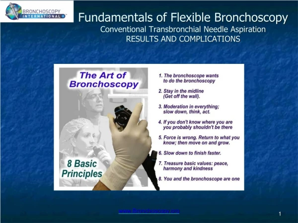
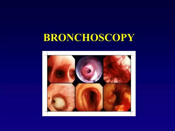
BACKGROUND • Allows direct visualization of the airways • Basic purposes: • Therapeutic (lavage and suction) • Diagnostic (inspect/collect samples for Dx) • Difficult intubation • Two types: • Rigid (under heavy sedation, removal of FBAO, surgical procedures) • flexible instruments (common, BAL…)
Bronch Steps • http://www.youtube.com/watch?v=XafLSohtOuk&feature=related • http://www.youtube.com/watch?v=hEA0TWEw5jU&feature=related • http://www.youtube.com/watch?v=AHNK_bs6YZU&feature=related
RIGID BRONCHOSCOPY • Generally performed by ENT’s and surgeons • Procedure oriented • Foreign body removal • Biopsies • Granuloma/polyp removal • Laser • Stent placement • Visualization for future surgery
FLEXIBLE BRONCHOCSOPY • Examination of the entire respiratory anatomy, nose to bronchi • Minor impact on anatomy • Able to pass through an endotracheal tube or tracheostomy tube
INDICATIONS • When flexible bronchoscopy is the best, easiest, safest, most efficient way to obtain the information
Bronch Assisting • Obtain the following medications and fluids: • 1 bottle of sterile normal saline 250 ml or greater. • Xylocain jelly for the scope • (2) bottles of 1% lidocaine (you will likely use only one) • At least 4 ml of sterile 4% lidocaine • Hurricane spray to numb the throat, may also have patient gargle lidocain, or inject it into trachea, may also use Cocaine. • 1 mg/ml epinephrine for possible bleeding • RN will administer sedative (Ativan or Versed usually)
Bronch Assisting • Setting up room Depending on setting (bedside or in procedure room, may also be done under fluoroscopy) • Have several 10-12 ml syringes for injection ready • Open saline bottle, have ready for injection and lavage • Open small specimen cups and lable 1% and 2-4% Lidocaine, have ready for injection • Have slides and specimen container ready for biopsies • Have cytology brush and forceps ready (keep sterile) • Have Leukins trap ready
Bronch Assisting • Setting up room • Gauze • Towels • Sterile gloves, gowns and masks, face shields • Lead vests if done in Fluoroscopy • Bite block • Suction equipment/tubing • Oxygen and resusitation bag
Bronch Assisting • When drawing up fluid for injection through the injection port of the bronchoscope, draw up several cc of air on top of the fluid. The air will follow the fluid through the bronchoscope to clear the channel of fluid. Fluid injected into the airway should be sterile • Obtain several patient labels to attach to laboratory specimens (you will need to write the sample type and site on the label (e.g. "Right upper lobe BAL", "bronchial washing", etc.) once obtained. Have available any laboratory forms that might be necessary for proper specimen processing (pathology, cytology, microbiology, etc).
Bronch Assisting • Organize at the bedside at least: 1 sterile biopsy forceps for bronchoscopy (have formalin available and a separate small container of sterile NS for rinsing the forceps if it touches the formalin). Assure smooth functioning of the forceps. • 1 sterile cytology brush for bronchoscopy (have several slides available with slide container and fixative – you will need to ask the physician if the slides should be "fixed" if you obtain a brushing. • 1 sterile transbronchial aspiration needle for bronchoscopy (have a 20-35 ml syringe to apply suction to the needle (assure it is compatible with the syringe port on the needle) and several slides available). • Compatibility of the external diameter of all scope accessories with the internal diameter of the bronchoscope should be verified before the procedure.
Bronch Assist • Assure the bronchoscope and other reusable items have been properly cleaned and disinfected. If the suction valve is reusable inspect it for possible debris left behind after cleaning. Plug the bronchoscope into the light source and "white balance" it (shine the tip at something white and push the "white balance" button) the look through it and assure it is in proper working order. Check any cameras and/or video equipment that may be used. Connect suction tubing to bronchoscope (during the procedure you will likely be using full suction vacuum setting) with an in-line suction trap (have at least 3 more suction traps available). Assure proper resuscitation equipment is in the bronchoscopy area.
Patient Prep • Obtain and review patient chart and x-rays then make them available for the physician performing the procedure. Patients typically are assessed for potential bleeding problems, etc - obtain any pre-procedure laboratory results (coagulation assessment, ECG, spirometry, etc). Record how long the patient has been NPO. Assess the ability to adequately oxygenate the patient during the procedure. Assess the patient for tuberculosis risk, as procedure may need to take place in specially ventilated room.
Patient Prep • Assure proper consent has been obtained. Obtain medication allergy and hypersensitivity information. Explain procedure to patient. Obtain and record baseline room air oximetry (if patient normally uses oxygen, obtain oximetry reading on their normal level of supplemental oxygen). Obtain and record baseline pulse reading. Obtain and record baseline blood pressure reading. Connect patient to ECG monitor.
Post Procedure • Monitor the following and record items as significant and vital signs on a regular basis: • Level of consciousness. • Medications administered, dosage, route, and time of delivery. • Subjective responses (e.g., pain, discomfort, dyspnea). • Blood pressure, heart rate, rhythm, and changes in cardiac status • SpO2 and supplemental oxygen use. • Patient should be observed until stable. • Patient should remain NPO for 2 hours and after this period has expired begin by trying small sips of water to assure the ability to effectively swallow. • Outpatients should be instructed to contact the bronchoscopist regarding fever, chest pain or discomfort, dyspnea, wheezing, hemoptysis, or any new findings presenting after the procedure has been completed. Oral instructions should be reinforced by written instructions that include names and phone numbers of persons to be contacted in emergency
TECHNIQUE • Anesthesia • Best accomplished in the operating room • May be performed bedside in an ICU setting • Continuous monitoring • Light anesthesia--allows continued spontaneous breathing • May be done with conscious sedation in older individuals
TECHNIQUE • Insertion • Nasal • LMA • Endotracheal tube • Tracheostomy tube • Appropriate topical anesthesia and lubrication
TECHNIQUE • Anatomical survey • Nasal passages • Pharynx • Larynx • Trachea • Bronchi • Examine all before any other procedures
TECHNIQUE • Additional procedures • Bronchoalveolar lavage • Brushings • Bronchial biopsy • Transbronchial biopsy • Laser • Others: cryotherapy, stent placement, foreign body removal, needle biopsy
BRONCHOALVEOLAR LAVAGE • Small aliquots of sterile normal saline instilled into the airway • Removed by suctioning • Samples distal bronchial and alveolar surfaces • Wedge position to prevent loss of fluid
BAL TESTS • Microbiology • Bacterial, viral, fungal, AFB, special techniques • Pathology • Cell count, differential, special stains
MICROBIOLOGIC STUDIES • Stains • Gram stain • Acid fast stain (Ziehl-Neelsen) • Antibody tests • Rapid tests, DFA tests (direct fluorescent antibody (DFA) testing) Ex: RSV • In-situ (tumors) • PCR (polymerase chain reaction) DNA
SPECIAL STAINS • Fungi • Silver (Gomori’s methenamine silver stain) • Pneumocystis carinii • Silver stain • Papanicolaou
SPECIFIC INDICATIONS • Atelectasis • Recurrent pneumonia • Chronic cough • Persistent/unexplained wheeze • Hemoptysis • Suspected airway compression/obstruction • Stridor • Upper airway obstruction • Suspected aspiration • Evaluation of tracheostomies
Lung Cancer in Bronchus http://www.youtube.com/watch?v=ezsD0OjqbH8
Stenosis Post Laser Surgery
Tracheal Cleft http://www.youtube.com/watch?v=Po208uGAeww tracheal clefts and congenitally short tracheas