Download
1 / 32
330 likes | 379 Views
Chapter 9 Intraocular Lenses. Which spectacle-corrected 4 D anisometrope would have the greatest problems with accommodative imbalance in near vision?. 45 year old wearing PALs with +1.25 D maximum add power 65 year old wearing bifocals with +2.50 D near add power

E N D
Which spectacle-corrected 4 D anisometrope would have the greatest problems with accommodative imbalance in near vision? • 45 year old wearing PALs with +1.25 D maximum add power • 65 year old wearing bifocals with +2.50 D near add power • 40 year old with single vision distance correction • 25 year old with single vision distance correction
All pre-presbyopic spectacle-corrected astigmats have imperfect near vision. What is the basis of their problem? • Reduced effective cylinder at near • Excess effective cylinder at near • Rotation of principal meridians at near • Insufficient ocular amplitude of accommodation
Which of the following uncorrected hyperopes would have the greatest problem with distance vision? • Manifest hyperopia 4 D, facultative hyperopia 4 D, latent hyperopia 0.5 D • Latent hyperopia 1.5 D, absolute 0 D • Latent hyperopia 0.5 D, total 4 D, facultative 3 D • Total hyperopia 6 D, facultative hyperopia 4 D, latent 2 D
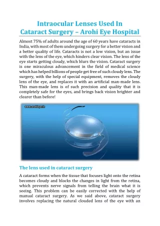
Page 9.1 Cataracts and Intraocular Lenses • After cataract extraction, the emmetropic eye becomes about 12 D hyperopic • Before the days of IOLs, patients were prescribed high plus spectacle lenses or even higher plus contacts • IOLs (first used in the 1960s) replace the crystalline lens and are designed to make the patient emmetropic
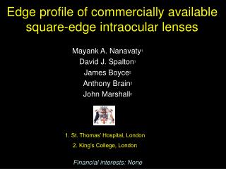
Page 9.1 Aphakic IOL Implantation • Earliest cataract extraction procedures were Intracapsular (removal of entire crystalline lens and capsule). Required large incision. • Older IOLs inflexible (e.g. PMMA), so larger incision was also required for implant insertion • Larger incision often led to significant (and variable) post-surgical astigmatism and other complications • Most cataract extractions today are Extracapsular (capsule remains intact). Smaller incision required • Newer IOL designs are foldable and can fit through the smaller incisions that are possible with extracapsular extraction
Extracapsular Cataract Extraction Used when lens nucleus too dense for emulsification
Early Anterior Chamber IOLs (1960s) Fig 9.1 Page 9.1 • Intracapsular cataract extraction • PMMA “Iris Clip” Lens placed in anterior chamber • Many iris-related problems: iritis, pupil distortion, corneal endothelial cell loss
Posterior Chamber IOLs (1977) Fig 9.2 Page 9.2 • Extracapsular cataract extraction (capsule intact) introduced in late 1960s • Allowed posterior chamber implantation, initially in ciliary sulcus • Capsular bag soon took over as implant site of choice because of problems with ciliary sulcus implants (e.g. pigmentary glaucoma)
Fig 9.3 Page 9.2 Ciliary Sulcus
Fig 9.4 Page 9.3 Posterior Chamber IOL in Ciliary Sulcus
Sutured Haptic tied off and knot buried in conjunctiva Fig 9.5 Page 9.3
Fig 9.6 Page 9.4 Capsular Bag Implant
Fig 9.7 Page 9.4 IOL inside Capsular Bag
Staar Lens Implanted in Capsular Bag Staar Lens (foldable silicone) Fig 9.8 Page 9.4 Newer Capsular Bag Lenses
Page 9.5 Phakic IOLs • Emergence of phakic IOLs in mid-1980s, as biocompatible foldable materials became available • Phakic IOL designs prior to 2000 excluded the ciliary sulcus and capsular bag as implant sites • AC IOLs therefore returned • Had to overcome the previous iris-related problems with AC lens • Advantage over LASIK, PRK etc. reversible
Fig 9.9, Page 9.5 • Iris claw lens (Artisan, 1998) • Not feasible with AC depth < 3.2 mm • This impacted primarily the hyperopic pool • Unfortunate because hyperopes have lower success rate with corneal refractive surgery than myopes • Complications (e.g. endothelial cell loss, glare, etc.) remain but appear to be decreasing with newer designs Lens attached to iris in mid-peripheral stroma
Fig 9.10 Page 9.6 Posterior Chamber Phakic IOLs • “Collamer” posterior chamber phakic ICL (implantable contact lens) • Implanted between iris and anterior crystalline lens • Contact with anterior lens causes anterior subcapsular cataract • Iris problems also occur • Best option for hyperopes • PC location means higher lens power than “equivalent corneal power” change with LASIK
Page 9.7 IOLs and near Vision • Multifocal intraocular lenses are the IOL equivalent of multifocal contact lenses • Poor track record until recently
Fig 9.12 Page 9.8 “Array” Multifocal Lens • Alternating distance and intermediate/near zones
Fig 9.13 Page 9.8 Accommodating IOLs • “Humanoptics” aphakic IOL • Capsular bag-fixated lens • Four flexible haptics that bend when constricted by capsular bag • Effect forward translation of lens • This increases total ocular power
Humanoptics Accommodating IOL unaccommodated accommodated