Download
1 / 42
440 likes | 701 Views
VIRAL MYOCARDITIS AN UPDATE. BY JAMEEL A. ALATA, MD. CONSULTANT & ASSISTANT PROFESSOR OF PEDIATRICS & PEDIATRIC CARDIOLOGY. INTRODUCTION.

E N D
VIRAL MYOCARDITIS AN UPDATE BY JAMEEL A. ALATA, MD. CONSULTANT & ASSISTANT PROFESSOR OF PEDIATRICS & PEDIATRIC CARDIOLOGY
INTRODUCTION • Myocarditis is an inflammatory disorder of the myocardium with necrosis of the myocytes and associated inflammatory infiltrate. It is usually caused by a viral infection, particularly adenovirus and enterovirus infections (eg, coxsackievirus) • suspected myocarditis can be classified into the following 3 types based on pathologic findings as defined in the Dallas Criteria (1987)
Active myocarditis is characterized by abundant inflammatory cells and myocardial necrosis. • Borderline myocarditis is characterized by an inflammatory response, but the inflammatory response is too sparse for this type to be labeled as active myocarditis. Degeneration of myocytes is not demonstrated by light microscopy. • Nonmyocarditis • If an active or borderline inflammatory process is found, follow-up biopsies can be subclassified into ongoing, resolving, or resolved myocarditis.
Pathophysiology • Myocarditis generally results in a decrease in myocardial function, with concomitant enlargement of the heart and an increase in the end-diastolic volume caused by increased preload. • The progressive increase in left ventricular end-diastolic volume increases left atrial, pulmonary venous, and arterial pressures, resulting in increasing hydrostatic forces. These increased forces lead to both pulmonary edema and congestive heart failure.
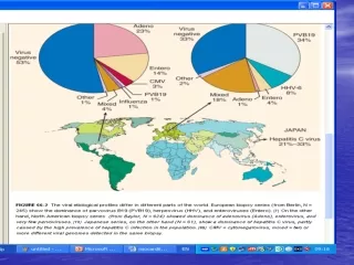
Frequency • The World Health Organization reports that incidence of cardiovascular involvement after enteroviral infection is 1-4%, • Incidence varies greatly among countries and is related to hygiene and socioeconomic conditions. Availability of medical services and immunizations also affect incidence.
Occasional epidemics of viral infections have been reported with an associated higher incidence of myocarditis. • Enteroviruses, such as coxsackievirus and echovirus, and adenoviruses, particularly types 2 and 5, are the most commonly involved organisms.
Mortality/Morbidity • With suspected coxsackievirus B, the mortality rate is higher in newborns (75%) than in older infants and children (10-25%). • Complete recovery of ventricular function has been reported in as many as 50% of patients. • Some patients develop chronic myocarditis (ongoing or resolving) and/or dilated cardiomyopathy and may eventually require cardiac transplantation.
Epidemiology • No racial predilection exists. • No sex predilection exists in humans, but there is some indication in laboratory animals that the disease may be more aggressive in males than in females. • Certain strains of female mice had a reduced inflammatory process when treated with estradiol.
In other studies, testosterone appeared to increase cytolytic activity of T lymphocytes in male mice. • No age predilection exists. • Younger patients, especially newborns and infants, and immunocompromised patients may be more susceptible to myocarditis.
CLINICAL • Heart failure: This is the most common presenting picture in all ages. • Chest pain: Although rare in young children, this may be the initial presentation for older children, adolescents, and adults. • Chest pain may be due to myocardial ischemia or concurrent pericarditis.
Arrhythmia: Patients can present with any type of dysrhythmia, including ; 1 ) Atrioventricular conduction disturbances. 2 ) Sinus tachycardia is typical and the rate is faster than expected for the degree of fever present, which is typically low-grade. 3 )Junctional tachycardia is also seen and can be difficult to control medically.
Dilated cardiomyopathy: • There is still debate over whether myocarditis progresses to dilated cardiomyopathy. • Many investigators believe that dilated cardiomyopathy is a direct result of a previously burned-out myocarditis episode.
Initial symptoms in infants include the following: • Irritability • Lethargy • Periodic episodes of pallor • Fever
Hypothermia • Tachypnea • Anorexia • Failure to thrive
Physical EXAM • Tachycardia, weak pulse, cool extremities, decreased capillary refill, and pale or mottled skin may be present. • Heart sounds may be muffled, especially in the presence of pericarditis. An S3 may be present, and a heart murmur caused by atrioventricular valve regurgitation may be heard.
Hepatomegaly may be present in younger children. • Rales may be heard in older children. • Jugular venous distention and edema of the lower extremities may be present.
Neonates • Neonates may seem irritable, be in respiratory distress, and exhibit signs of sepsis. • Somnolence, hypotonia, and seizures can be associated if the CNS is involved. • Hypothermia or hyperthermia, oliguria, elevated liver enzymes and elevated blood urea nitrogen and creatinine caused by direct viral damage and/or low cardiac output may be present.
Infants • Signs include failure to thrive, anorexia, tachypnea, tachycardia, wheezing, and diaphoresis with feeding. • In severe cases, low cardiac output may progress to acidosis and death. • End organ damage may occur because of direct viral infestation or because of low cardiac output. • CNS involvement may also occur.
Adolescents • Presentation may be similar to that of younger children but with a more prominent decrease in exercise tolerance, lack of energy, malaise, chest pain, low-grade fever, arrhythmia, and cough. • End-organ damage and low cardiac output may be present.
Causes Infecting organisms include the following: • Coxsackievirus types A and B, especially type B, are the most common viral causes of myocarditis. • Adenovirus (types 2 and 5 most common) • Cytomegalovirus • Echovirus • Epstein-Barr virus • Hepatitis C virus
Herpes virus • Human immunodeficiency virus • Influenza and parainfluenza • Measles • Mumps, associated with endocardial fibroelastosis (EFE) • Parvovirus B19 • Poliomyelitis virus • Rubella • Varicella
Murine model • The coxsackievirus and adenovirus receptor acts as the receptor for the four most common viruses causing human myocarditis: Type C (type 2 and type 5) adenovirus and Coxsackievirus B3 and B4. • Coxsackievirus B serotypes 1-6 have been associated with human myocarditis, but the most serious cases have been attributed to types 3 and 4.
PATHOPHYSIOLOGY • The primary response to the early phase of viral infection is the release of natural killer (NK) cells, which lyse infected myocytes. This helps clear the virus from the system.
NK cells also induce expression of major histocompatibility complex antigens on myocytes by releasing cytokines, which prepare the NK cells to interact with T lymphocytes. • Animal models depleted of NK cells develop a more severe form of myocarditis.
The late phase or second wave of T lymphocytes (CD4, CD8) begins approximately 1 week after the mouse has been inoculated with the virus. T lymphocytes can injure cells in the following 3 ways: • Stimulation of cytotoxic T cells • Production of antibody and antibody-dependent myotoxicity • Direct antibody and complement formation
These ongoing processes are considered genetically mediated autoimmune processes. • Two different strains of cytolytic T cells have been recognized; one strain attacks virus-infected myocytes and the other strain attacks uninfected cells. • Enzymatic cleavage by viral proteins of cytoskeletal proteins appears to play a role in development of dilated cardiomyopathy.
Apoptosis appears to play a role also in the development of dilated cardiomyopathy. • Various kinds of autoantibodies have been found in as many as 60% of patients with myocarditis.
These include • complement-fixing antimyolemmal antibodies, • complement-fixing antisarcolemmal antibodies, • antimyosin heavy chain antibodies, and • anti–alpha myosin antibodies. Although their role in the disease is not completely understood, their presence may serve as a marker for diagnosing myocarditis in the future.
DIFFERENTIALS • Anomalous Left Coronary Artery from the Pulmonary Artery. • Aortic Stenosis, Valvar • Cardiac Tumors • Cardiomyopathy, Dilated • Carnitine Deficiency • Coarctation of the Aorta
Coronary Artery Anomalies • Endocardial Fibroelastosis • Enteroviral Infections • Glycogen Storage Disease Type I • Glycogen Storage Disease Type II • Myocarditis, Nonviral • Pericarditis, Viral
Investigations • Virus identification ; 1 ) Cultures from blood , stools and throat. 2 ) Acute & convalescent sera.
ECG • CXR • Echocardiogram
CBC • PT , PTT , FDP , & D-diamers • ABG • LFT
Renal function • Cardiac enzymes • Carnitine level
Treatment • Bedrest or limitation of activity in the acute phase. • Intubation & ventilation. • Treatment of PHTN • Diuretics. • Inotropes
Digoxin ( better no loading & later on in the course of the disease ). • High dose IVIG. • ACE inhibitors. • Corticosteroids ? • Sedation and paralysis.
[Clinical study on therapeutic effects of treatment according to syndrome differentiation of traditional Chinese medicine combined with captopril on severe viral myocarditis complicated heart failure]( CHINESE ) • RESULTS: The therapeutic effect of the treated group according to NYHA classification was obviously better than that of the control group. • The creatine phosphokinase isoenzyme (CPK-MB), aspartate transaminase (AST), lactate dehydrogenase (LDH) content lowered in both groups, but more significantly lowered in the treated group than in the control group (P < 0.05, P < 0.01). • The improvement of S-T segment of ECG in the treated group was better than that in the control (P < 0.01); also some parameters of heart function and motorial toleration were bettered in the treated group more significantly (P < 0.01).
Carvedilol increases the production of interleukin-12 and interferon-gamma and improves the survival of mice infected with the encephalomyocarditis virus.( JAPAN ) • RESULTS: • Carvedilol 1)Improved the 14-day survival of the animals. 2)Attenuated myocardial lesions on day 7, and 3 )Increased myocardial levels of interleukin (IL)-12 and interferon (IFN)-gamma, whereas reducing myocardial virus replication. • Propranolol also attenuated myocardial lesions, but to a lesser extent, and increased IL-12 and IFN-gamma levels. • Metoprolol had no effect in this model. Encephalomyocarditis virus infection increased plasma catecholamine levels.
Successful treatment of enterovirus-induced myocarditis with interferon-alpha.( ITALY ). • Non- randomized, placebo-controlled studies have investigated interferon-alpha therapy in enterovirus-proven myocarditis. • This report describes 2 patients with enterovirus-induced myocarditis (1 with associated Churg-Strauss syndrome) who at follow-up endomyocardial biopsy showed clinical and hemodynamic improvement and viral clearance (using polymerase chain reaction) after interferon-alpha therapy.
Cardiac MRI in suspected myocarditis ( GERMANY ) • Acute myocarditis was diagnosed in 9 patients and cardiac sarcoidosis in 2 patients. Late enhancement was observed in 4 patients with acute myocarditis and in both patients with cardiac sarcoidosis. • Semiquantitative evaluation revealed 9 true positive, 9 true negative, 1 false positive and 2 false negative results. • CONCLUSION: Cardiac MRI has the potential to detect acute myocarditis and to diagnose cardiac sarcoidosis. Late enhancement of Gd-DTPA can be found in both viral myocarditis and cardiac sarcoidosis.
Here we show the essential role of Janus kinase (JAK) signaling in cardiac myocyte antiviral defense and a negative role of an intrinsic JAK inhibitor, the suppressor of cytokine signaling (SOCS), in the early disease process. ( USA ) • strategies directed at inhibition of SOCS in the heart and perhaps other organs can augment the host-cell antiviral system, thus preventing viral-mediated end-organ damage during the early stages of infection.