Download
1 / 96
1.31k likes | 2.48k Views
colorectal cancer. By Dr. Ryan Al.Ghanemi. overview of the lecture. case presentation. epidemiology of colorectal cancer. clinical presentation of colorectal cancer. staging of colorectal cancer. management of primary colon tumor. management of rectal tumo r. case presentation.

E N D
colorectal cancer • By Dr. Ryan Al.Ghanemi
overview of the lecture • case presentation. • epidemiology of colorectal cancer. • clinical presentation of colorectal cancer. • staging of colorectal cancer. • management of primary colon tumor. • management of rectal tumor.
case presentation • A 65 years old man present with dyspnea on exertion and angina for 3-4 weeks. • He denies any cough, weight loss, git symptoms. • His past medical history is significant for hypertension, stable angina, and colonic polyps • Those were removed 7 to 8 years ago by colonoscopy.
The physical examination reveals a well-nourished man. • The finding from head and neck, cardiopulmonary, and neurologic examinations are Unremarkable. • Examination of the abdomen reveals an obese abdomen without tenderness or Palpable masses . • The rectal examination reveals no masses , a smooth enlarged prostate
Strongly hemoccult positive stool in the vault . • Cbc : hb of 8.7 g/dl hct 29 % and low mcv . • Ecg : normal sinus rhythm and mild lvh. • Normal chest radiograph.
What is the most likely mechanism causing this process? • How would you confirm the diagnosis ? • What is the initial treatment for this pt ?
Most likely mechanism: anemia caused by occult GI tract bleeding . • Confirmation of the diagnosis : EGD and colonscopy. • Initial treatment: blood transfusion.
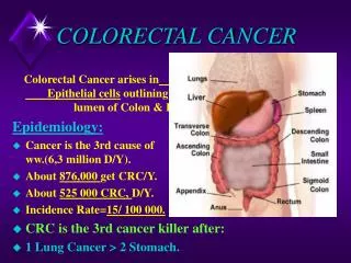
Epidemiology: • Approximately 148,810 new cases of large bowel cancer are diagnosed each year in the United States, of which 108,070 are colon and the remainder rectal cancers. • Annually, approximately 49,960 Americans die of CRC, accounting for approximately 9 percent of all cancer deaths.
Incidence • Age is a major risk factor for sporadic CRC. It is a rare diagnosis before the age of 40, the incidence begins to increase significantly between the ages of 40 and 50, and age-specific incidence rates increase in each succeeding decade thereafter).
The lifetime incidence of CRC in patients at average risk is about 5 percent, with 90 percent of cases occurring after age 50. • The incidence is higher in patients with specific inherited conditions that predispose them to the development of CRC.
A gradual shift toward right-sided or proximal colon cancers has been observed both in the United States and internationally. • The greatest increase in incidence is in cecal primaries.
why this changes? • This change in the anatomic distribution of CRCs may be, in part, related to improvements in diagnosis and treatment. • and increased screening by flexible sigmoidoscopy with removal of adenomatous polyps in the descending colon . • but there also appears to be a true increase in the incidence of ascending colon and cecal cancers .
Consistent with this hypothesis, five-year survival rates have improved significantly for left and transverse colon cancers, but not for right-sided tumor.
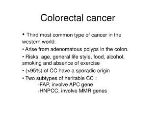
Risk factors : • Environmental and genetic factors can increase the likelihood of developing CRC. • Although inherited susceptibility results in the most striking increases in risk. • the majority of CRCs are sporadic rather than familial .
Personal or family history of sporadic cancers or adenomatous polyps — Patients with a personal history of CRC or adenomatous polyps are at risk for the development of a future large bowel cancer
FAP • Hereditary nonpolyposis colorectal cancer • A personal history of large (>1 cm) adenomatous polyps and polyps with villous or tubulovillous histology also increase the risk of CRC, particularly if multiple . The relative risk ranges from approximately 3.5 to 6.5 in such patients
Family history is also an important risk factor in sporadic disease, with a single affected first-degree relative (parent, sibling, or child) increasing the risk 1.7-fold of that in the general population • A family history of a large (>1 cm) or histologically advanced colonic adenoma appears to carry the same significance as a positive family history of colorectal cancer
Inflammatory bowel disease • Diabetes mellitus and insulin resistance obesity • Cholecystectomy - Alcohol • A history of radiation therapy for prostate cancer was associated with an increased risk of rectal cancer in a large database study . The magnitude of risk was similar to that observed in patients with a family history of colonic adenomas
protective factors • Diet • Fiber • Calcium supplementation has been recommended for the primary or secondary prevention of colonic adenomas by the American College of Gastroenterology
Aspirin and NSAIDs • Hormone replacement therapy
Omega 3 fatty acids — Consumption of omega 3 fatty acids (mainly as fish oil) has been associated with a reduced incidence of colorectal cancer in observational studies
Screening: • Patient surveillance can most effectively be accomplished by colonoscopy. • For average risk individuals ACG colonoscopy every 10 years beginning at 50 years of age . • If adenomatous polyp is identified and removed repeat colonoscopy should be done every 3 years when the colon is clear every 5 years . • What if he has high risk factor , eg early family history ?
Clinical presentations: • • Abdominal pain — 44 percent • • Change in bowel habit — 43 percent • • Hematochezia or melena — 40 percent • • Weakness — 20 percent • • Anemia without other gastrointestinal symptoms — 11 percent • Weight loss — 6 percent
A meta-analysis of 15 studies concluded that the sensitivity of alarm features (such as weight loss) was poor (ranging from 5 to 64 percent). • However, the specificity for some alarm symptoms (including dark red rectal bleeding and abdominal mass) was greater than 95 percent.
Abdominal pain can be caused by a partial obstruction, peritoneal dissemination, or intestinal perforation leading to generalized peritonitis. • tenesmus caused by a rectal cancer may involve pelvic floor muscles. • a locally advanced lesion may involve the sciatic or obturator nerve, leading to a neuropathic pain syndrome
Hematochezia is more often caused by a rectal rather than colon cancer • A change in bowel habits is a more common presenting symptom for left-sided cancers because fecal contents are liquid in the proximal colon and are therefore less likely to be associated with obstructive symptoms
Metastatic disease — Approximately 20 percent of patients have distant metastatic disease at the time of presentation • The presence of right upper quadrant pain, abdominal distention, early satiety, supraclavicular adenopathy, or periumbilical nodules usually signals advanced disease.
Because the venous drainage of the intestinal tract is via the portal system, the first site of hematogenous dissemination is usually liver • tumors arising in the distal rectum may metastasize initially to the lungs because the inferior rectal vein drains into the inferior vena cava rather than into the portal venous system.
Unusual presentations — There are also a variety of unusual presentations of CRC. These include: • Local invasion or a contained perforation causing malignant fistula formation into adjacent organs • Fever of unknown origin • CRC ultimately proves to be the origin of approximately 6 percent of adenocarcinomas of unknown primary sites
Impact of symptoms on prognosis • — The presence of symptoms and their particular type appear to be of some prognostic importance:
• Patients who are symptomatic at diagnosis have a somewhat worse prognosis . In one report, the five-year survival rate for symptomatic and asymptomatic patients was 49 versus 71 percent. • the duration of symptoms is not an accurate predictor of prognosis.
• Obstruction and/or perforation, although uncommon, carry a poor prognosis, independent of stage . • • Tumors presenting with hemorrhage have been thought to have a better prognosis because of their tendency to be diagnosed earlier; however, bleeding is not an independent predictor of outcome .
LOCATION OF COLORECTAL MALIGNANCIES • Synchronous cancers — Synchronous CRCs: • defined as two or more distinct primary tumors separated by normal bowel and not due to direct extension or metastasis, occur in 3 to 5 percent of patients with colon cancer
Metachronous cancers : • Metachronous CRCs, defined as nonanastomotic new tumors developing at least six months after the initial diagnosis. • develop in 1.5 to 3 percent of patients in the first five years postoperatively, rising to up to 9 percent after several decades in survivors of the primary cancer
DIAGNOSIS • CRC may be suspected from one or more of the symptoms and signs described above or may be asymptomatic and discovered by routine screening of average and high risk subject
Colonoscopy and BE • The vast majority of colon and rectal cancers are endoluminal adenocarcinomas that arise from the mucosa. Colonoscopy is the single best diagnostic test in symptomatic individuals, since it can localize lesions throughout the large bowel, biopsy mass lesions, detect synchronous neoplasms, and remove polyps • The air contrast barium enema (BE), supplemented with flexible sigmoidoscopy, is also used to evaluate symptomatic patients
A direct evaluation of the total colonic mucosa is necessary to exclude carcinoma with certainty
staging • Tx: No description of the tumor's extent is possible because of incomplete information. • Tis: The cancer is in the earliest stage. It involves only the mucosa. It has not grown beyond the muscularis mucosa (inner muscle layer). • T1: The cancer has grown through the muscularis mucosa and extends into the submucosa. • T2: The cancer has grown through the submucosa and extends into the muscularis propria (outer muscle layer).
T3: The cancer has grown through the muscularis propria and into the subserosa but not to any neighboring organs or tissues. • T4: The cancer has grown through the wall of the colon or rectum and into nearby tissues or organs.
N categories for colorectal cancer • N categories indicate whether or not the cancer has spread to nearby lymph nodes and, if so, how many lymph nodes are involved. • Nx: No description of lymph node involvement is possible because of incomplete information. • N0: No lymph node involvement is found. • N1: Cancer cells found in 1 to 3 nearby lymph nodes. • N2: Cancer cells found in 4 or more nearby lymph nodes.
M categories for colorectal cancer • M categories indicate whether or not the cancer has spread to distant organs, such as the liver, lungs, or distant lymph nodes. • Mx: No description of distant spread is possible because of incomplete information. • M0: No distant spread is seen. • M1: Distant spread is present.
Stage 0 • Tis, N0, M0: The cancer is in the earliest stage. It has not grown beyond the inner layer (mucosa) of the colon or rectum. This stage is also known as carcinoma in situ or intramucosal carcinoma. • Stage I • T1, N0, M0 or T2, N0, M0: The cancer has grown through the muscularis mucosa into the submucosa (T1) or it may also have grown into the muscularis propria (T2). It has not spread to nearby lymph nodes or distant sites.
Stage IIA • T3, N0, M0: The cancer has grown into the outermost layers of the colon or rectum but has not reached nearby organs. It has not yet spread to the nearby lymph nodes or distant sites. • Stage IIB • T4, N0, M0: The cancer has grown through the wall of the colon or rectum and into other nearby tissues or organs. It has not yet spread to the nearby lymph nodes or distant sites.
Stage IIIA • T1, N1, M0 or T2, N1, M0: The cancer has grown through the mucosa into the submucosa (T1) or it may also have grown into the muscularis propria (T2). It has spread to 1 to 3 nearby lymph nodes but not to distant sites. • Stage IIIB • T3, N1, M0 or T4, N1, M0: The cancer has grown into the outermost layers of the colon or rectum but has not reached nearby organs (T3) or the cancer has grown through the wall of the colon or rectum and into other nearby tissues or organs (T4). It has spread to 1 to 3 nearby lymph nodes but not distant sites.
Stage IIIC • Any T, N2, M0: The cancer may or may not have grown through the wall of the colon or rectum, but it has spread to 4 or more nearby lymph nodes. It has not spread to distant sites. • Stage IV • Any T, Any N, M1: The cancer may or may not have grown through the wall of the colon or rectum, and it may or may not have spread to nearby lymph nodes. It has spread to distant sites such as the liver, lung, peritoneum (the membrane lining the abdominal cavity), or ovary.
An increase in colorectal cancer screening has been associated with an earlier stage at which colorectal cancer is diagnosed. The following results were observed in a large database study • • Localized — confined to the primary site and to the mucosa, submucosa, and muscle layer (Dukes' A or B or TNM stage I or II ) — 40 percent • • Lymph node involvement (Dukes' C or TNM stage III) — 37 percent • • Distant metastases (Dukes' D or TNM stage IV) — 19 percent
As a general rule, the stage of rectal cancer at diagnosis tends to be slightly earlier than the stage in the colon probably because rectal cancers are more likely to cause symptoms.
In patients with four or fewer hepatic lesions, resection may be curative, with five-year relapse-free survival rates of 24 to 38 percent