Download
1 / 23
350 likes | 975 Views
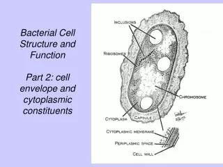
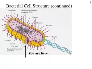
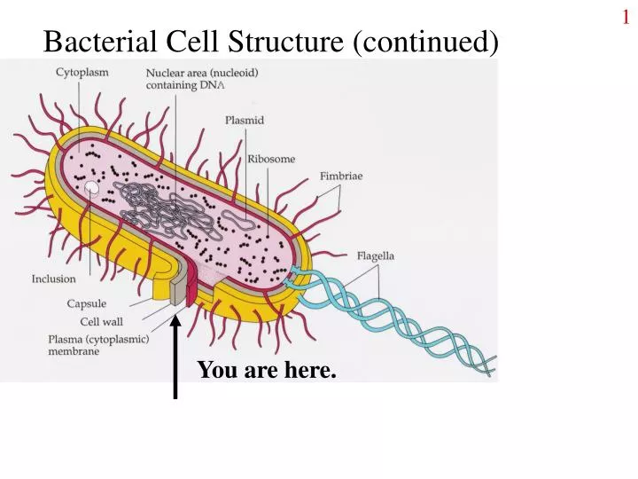
Bacterial Cell Structure (continued). You are here. Peptidoglycan structure. Bacteria typically face hypotonic environments Peptidoglycan provides support, Limits expansion of cell membrane Bacteria need other protection from hypertonic situations. Gram negative cell wall. Outer membrane.

E N D
Bacterial Cell Structure (continued) You are here.
Peptidoglycan structure • Bacteria typically face hypotonic environments • Peptidoglycan provides support, Limits expansion of cell membrane • Bacteria need other protection from hypertonic situations
Outer membrane • Lipid bilayer membrane • Inner and outer leaflets • Inner leaflet made of phospholipids; outer leaflet is made of lipopolysaccharide (LPS) • LPS = endotoxin • Proteins for transport of substances • Porins: transmembrane proteins • Barrier to diffusion of various substances • Lipoprotein: anchors outer membrane to PG
Structure of LPS extends from cell surface. contains odd sugars e.g. KDO. Gln-P and fatty acids take the place of phospholipids. www.med.sc.edu:85/fox/ cell_envelope.htm
Periplasmic Space www.arches.uga.edu/~emilyd/ theory.html
Periplasmic space: • A lot like cytoplasm, with • Peptidoglycan layer • Proteins that aid in transport • Proteins that break down molecules • Proteins that help in synthesis.
Glycocalyx: capsules and slime layers “Sugar covering”: capsules are firmly attached, slime layers are loose. Multiple advantages to cells: prevent dehydration absorb nutrients protection from predators, WBCs protection from biocides (as part of biofilms) attachment to surfaces and site of attachment by others. S-layers are highly structured protein layers that function like capsules cell capsule www.activatedsludge.info/ resources/visbulk.asp
Fimbriae and pili Both are appendages made of protein Singular: fimbria, pilus Both used for attachment Fimbriae: to surfaces (incl. host cells) and other bacteria. Pili: to other bacteria for exchanging DNA (“sex”). www.ncl.ac.uk/dental/oralbiol/ oralenv/images/sex1.jpg
Flagella • Flagella: protein appendages for swimming through liquid or across wet surfaces. • Rotate like propellers. • Different from eukaryotic flagella. • Arrangements on cells: • polar, • Lophotrichous, • amphitrichous, • peritrichous. www.ai.mit.edu/people/ tk/ce/flagella-s.gifwww.bmb.leeds.ac.uk/.../icu8/ introduction/bacteria.html
Prokaryotic vs. eukaryotic flagella • Prokaryotic flagella: • Made of protein subunits • Protrude through cell wall and cell membrane. • Stiff, twirl like a propeller • Eukaryotic flagella: • A bundle (9+2) of microtubules (made of protein) • Covered by cell membrane • Whipping action www.scu.edu/SCU/Departments/ BIOL/Flagella.jpg img.sparknotes.com/.../monera/ gifs/flagella.gif
Chemotaxis Bacteria change how they move in response to chemicals Bacteria move toward attractants (e.g. nutrients). Bacteria move away from repellants. In this figure, bacteria use up nutrients in the agar, then move outward to where more nutrients are, producing rings of growth. http://class.fst.ohio-state.edu/fst636/SP2004_mustafa/chemotaxis%20demo_SP04.htm
Runs and Tumbles: bacteria find their way http://www.bgu.ac.il/~aflaloc/bioca/motil1.gif
Spirochetes have internal flagella • Axial filament: a bundle of internal flagella • Between cell membrane and outer membrane in spirochetes • Filament rotates, bacterium corkscrews through medium Some bacteria move without flagella • Gliding • No visible structures, requires solid surface • Slime usually involved.
Axial filaments http://images.google.com/imgres?imgurl=http://microvet.arizona.edu/Courses/MIC420/lecture_notes/spirochetes/gifs/spirochete_crossection.gif&imgrefurl=http://microvet.arizona.edu/Courses/MIC420/lecture_notes/spirochetes/spirochete_cr.html&h=302&w=400&sz=49&tbnid=BOVdHqepF7UJ:&tbnh=90&tbnw=119&start=1&prev=/images%3Fq%3Daxial%2Bfilament%2Bbacteria%26hl%3Den%26lr%3D%26sa%3DG
Gliding Motility Movement on a solid surface. No visible organelles of locomotion. Cells produce, move in slime trails. Unrelated organism glide: myxobacteria, flavobacteria, cyanobacteria; appear to glide by different mechanisms. Cells glide in groups, singly, and can reverse directions. www.microbiology.med.umn.edu/ myxobacteria/trails.jpg
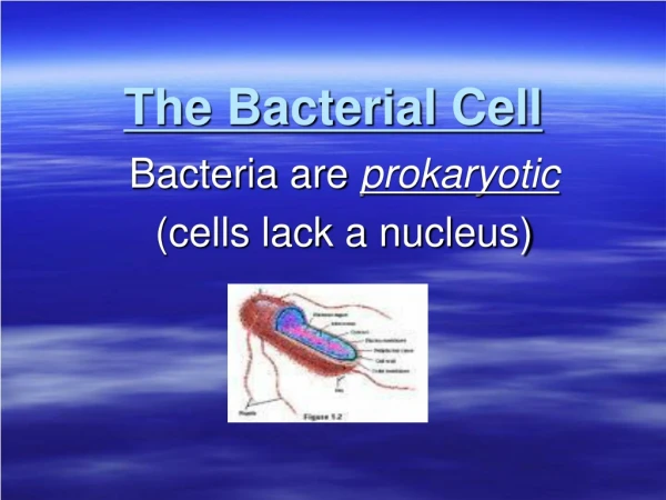
From the membrane in: the bacterial cytoplasm • Cytoplasm is a gel made of water, salts, LMW molecules, and lots of proteins. • DNA = nucleoid, w/ proteins • Plasmids = small circular DNA • Ribosomes: site of protein synthesis. Cytoplasm may also contain inclusions, gas vacuoles, extended membrane systems, or magnetosomes. But generally NO membrane-bound organelles.
Inclusions and granules • Storage molecules found as small bodies within cytoplasm. • Can be organic (e.g. PHB or glycogen) or inorganic (Sulfur, polyphosphate. • PHB, a type of PHA, degradable plastic (polyester); glycogen, a polymer of glucose. • Sulfur, a metabolic by-product; polyphosphate, polymer of PO4 www.qub.ac.uk/envres/EarthAirWater/ phosphate_removal.htm
Magnetosomes Membrane coated pieces of magnetite, assist bacteria in moving to microaerophilic environments. An organelle? North is down. Magnetospirillum magnetotacticum www.calpoly.edu/~rfrankel/ mtbphoto.html
How things get in (and out) of cells • Eukaryotic cells • Have transport proteins in membrane • Have a cytoskeleton made of microtubules • Allows for receptor mediated endocytosis, phagotcytosis, etc. • Cell membrane pinches in, creates vesicle • Prokaryotic cells • Have very little cytoskeleton • Can NOT carry out endocytosis • Entry of materials into cell by diffusion or transport processes ONLY.
Illustrations: entry into cells Both prokaryotes and eukaryotes. Only eukaryotes. http://bio.winona.msus.edu/bates/genbio/images/endocytosis.gif http://www.gla.ac.uk/~jmb17n/Teaching/JHteaching/Endocytosis/figures/howdo.jpg
How molecules get through the membrane Small molecules like gases can diffuse through the bilayer. Larger or more hydrophilic molecules require transport proteins. Active transport requires metabolic energy.
Review of eukaryotic cells Mitochondrion Plasmalemma (cell membrane)nucleus, ribosomeslysozomeendoplasmic reticulumgolgi body www.steve.gb.com/ science/cell_biology.html