Download

1 / 37
380 likes | 601 Views
Cutaneous and Urticarial Vasculitis. Roger W. Fox, M.D. Professor of Medicine/Pediatrics University of South Florida Division of Allergy and Immunology. Cutaneous Vasculitis. Rare, or uncommon clinical diagnosis

E N D
Cutaneous and Urticarial Vasculitis Roger W. Fox, M.D. Professor of Medicine/Pediatrics University of South Florida Division of Allergy and Immunology
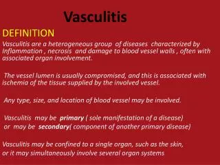
Cutaneous Vasculitis • Rare, or uncommon clinical diagnosis • Spectrum of presentation: clinical characteristics of cutaneousvasculitis may overlap with urticaria • Systemic diseases with cutaneousvasculitis constitute the majority of cases, but there are cases of cutaneousvasculitis without a systemic disorder • Constitutional symptoms often accompany the rash when associated with a systemic disorder • Laboratory tests and skin biopsy are diagnostic • Allergist/Immunologist most commonly are consulted to evaluate urticarialvasculitis
Urticarial Vasculitis:Clinical Features • Nonpruriticurticarial-like lesions and palpable purpura • Individual lesions persist 24-72 h (chronic urticaria lesions persist < 24 h) • Vasculitis is a rare cause of chronic urticaria (<5%) • Diagnosis: Skin biopsy and laboratory
Cutaneous VasculitisClassified • Small vessels(typical of urticarialvasculitis) and/or medium sized vessels (usual signs of medium size vasculitis are: purpura with necrosis, livedoreticularis, subcutaneous nodules, ulcerations, digital ischemia) • Inflammatory infiltrate on skin biopsy; mostly neutrophils, but eosinophils can be present • Direct immunofluorescencedermopathology assay: IgG, IgM, IgA and C3, fibrin deposition • Laboratory investigation: Complement studies, cryoglobulins, anti-neutrophil cytoplasmic antibodies (ANCA), ANA, CBC, ESR, serum protein electrophoresis c-ANCA, anti-proteinase 3 (PR3) associated with Wegener’s; p-ANCA, anti-myeloperoxidase (MPO) associated with Churg-Strauss
Vasculitis • Cutaneousvasculitis often presents as palpable purpuric lesions that may be generalized or limited to the lower extremities or other dependent areas. • Urticarial lesions, ulcers, and hemorrahagic blisters also occur. • May involve other organs liver, kidney, brain, and joints. Abbas K et al. Cellular and molecular immunology.6th Edition.
Direct Immunofluorescence Examination of a Skin-Biopsy Specimen. Kroshinsky D et al. N Engl J Med 2011;365:252-262
CutaneousVasculitis: Histopathology Criteria • Blood vessel damage (post-capillary venule in dermis) • Endothelial swelling • Fibrinoid necrosis • Leukocytoclasis (nuclear fragments) • Extravasation of RBCs • Perivascular inflammation • Complement components (immunoflorecence); immune complexes deposition
Cutaneous Vasculitis • Urticarialvaculitis- small vessels; C3, IgG, IgM* • Lupus vasculitis- small and medium-sized vessels; C3, IgG, IgM* • Cryoglobinemias- both size vessels; IgM+ • Hypersensitivity vasculitis (drug)- small vessel fibrin, C3+ • Polyarteritisnodosa (both, C3, IgG*), granulomatous polyangiitis (both, C3, IgG*) Henoch-Schonleinpurpura (both, IgA+) *perivascular and BMZ +perivascular
UrticarialVasculitis (UV) Differential Diagnosis • Chronic Idiopathic/Autoimmune Urticaria • Idiopathic UV • Systemic Diseases-SLE • Infections- Hepatitis B/C (cryoglobulinemia) • Serum Sickness and Drug Hypersensitivity Vasculitis • HypocomplementemicUrticarialVasculitis Syndrome (HUVS) • Churg-Strauss Syndrome • Schnitzler Syndrome- IgMgammopathy • Malignancies, immunologic and hematologic diseases reported
Serum Sickness • Chief manifestations 1 to 3 weeks after starting use of an offending agent. • Fever • Urticaria • Lymphadenopathy • Arthralgias Middleton’s. Allergy principles and practice. 7th Edition.
HypocomplementicUrticarialVasculitis Syndrome (HUVS) • Urticariavasculitis • Arthralgias/arthritis • Abdominal pain • Angioedema • Uveitis, scleritis, conjuncitivitis (unusual in SLE) • Proliferative glomerulonephritis (resembles SLE) • COPD, pleuritis(unusual in SLE) • Rarely, cardiovascular involvement: pericarditis, valvular disease • CNS involvement: aseptic meningitis, neuropathies
Clinical Photographs of the Patient. Kroshinsky D et al. N Engl J Med 2011;365:252-262
Skin-Biopsy Specimen from a Lesion (Hematoxylin and Eosin). Kroshinsky D et al. N Engl J Med 2011;365:252-262 neutophilic infiltration and leukocytoclasis
HUVS vs SLE • Granular deposition of immunoreactants along basement-membrane zone (characteristic of cutaneous lupus) • However, in conjunction with perivascularimmunoreactivity, suggests HUVS • Urticarial vasculitis and low complement levels are commonly associated with extra-cutaneous involvement as in SLE. HUVS has profoundly low complement levels
Vasculitis: Henoch-SchonleinPurpura • Small vessel vasculitis with IgA immune complexes • Classic triad of palpable purpura, arthritis, colicky abdominal pain. • Monitor for chronic nephritis • Usual course 4-6 weeks
CutaneousVasculitis • Drugs may be implicated in cutaneousvasculitis Direct immunofluorescent changes in these lesions suggest immune-complex deposition. • Drugs implicated: Allopurinol, thiazides, sulfonamides, other antimicrobials, several NSAIDs, interferons and anti-TNF α; metformin and sulfonylurea antidiabetic drugs. • The presence of eosinophils in the perivascular infiltrate of skin biopsy may indicate a higher probability of a drug etiology. http://emedicine.medscape.com/article/1049474-overview
Churg-Strauss Syndrome • “Allergic angiitis and granulomatosis” • Almost exclusively in individuals with asthma and allergic rhinitis; systemic vasculitis with eosinophilia, associated with fever, malaise, weight loss. • The pulmonary involvement dominates the clinical picture. Asthma is a defining feature and precedes the onset of vasculitis. Skin, heart, peripheral nervous system, GI tract and kidney can be involved.
CutaneousVasculitis:Laboratory • CBC with differential • Sedimentation rate • C-reactive protein • Complement studies • Autoantibody profiles • Liver and renal function • Serum protein electrophoresis
Treatments for CutaneousVasculitis • Corticosteroids • Hydroxychloroquine • Dapsone • Immunomodulating drugs
Schnitzler Syndrome • Onset as an adult (acquired disorder), although recently a gain of function mutation has been found in the NLRP3/CIAS-1 gene (1 patient) • Characterized by: • Recurring fevers • Neutrophilic urticaria • Monoclonal IgM gammopathy • Joint/bone pain • Similar presentation to MWS (no hearing loss) • May evolve into IgM multiple myeloma or Waldenstrom’s macroglobulinemia
Autoinflammatory Syndromes • Past 10 years • Newly identified classification of disease • Dysregulation of innate immunity • Recurrent fevers is common feature • Innate immunity recognize “danger" • Uncontrolled cytokine-mediated inflammation (IL-1B)
Muckle-Wells Syndrome (MWS) Moderate in severity of inflammation - more intense and long-lasting inflammation flares Characterized byurticarial rash with onset at birth, deafness and amyloidosis Sensorineural hearing loss Amyloidosis caused by build up of serum amyloid A (SAA) protein in kidneys which can lead to kidney failure Urticarial-like skin eruption in MWS Reprinted from Arth Rheum 2004 Feb; 50(2)607-612, Hawkins RN, et al. “Spectrum of Clinical Features of Muckle-Wells Syndrome and Response to Anakinra” with permission from Wiley InterScience. 33 Shinkai K, et al. Clin Exper Derm 2007;33:1-9.
NLRP3 Inflammasome pathway NLRP3 Inflammasome • The inflammasome is a large complex of proteins, the most important of which is NLRP31 • Mutations in the NLRP3 gene (a.k.a. CIAS1), which encode the cryopyrin (NALP3) protein, cause “autoinflammatory” diseases with a spectrum of onset and severity (MWS, FCAS)2,3 • Symptoms and pathology result from overexpression of Interleukin-13 *Ilaris is not FDA approved for NOMID NOMID = Neonatal Onset Multisystem Inflammatory Disease; MWS = Muckle Wells Syndrome; FCAS = Familial Cold Auto-Inflammatory Syndrome NLRP3= Nucleotide-binding oligomerization domain, leucine-rich-repeat family, pyrin domain containing 3 NALP3=NACHT leucine-rich repeat protein; CIAS1=Cold Induced Autoinflammatory Syndrome 1 34 1Neven B, et al. Nat Clin Pract Rheum 2008;4(9):481-489. 2Hoffman HM, et al. Arthr Rheum 2008;58(8):2443-2452. 3ChurchLD, et al. Nat Clin Pract Rheum 2008;4(1):34-42.
Urticarial lesions on trunk (A). Within 24 hours of commencing anakinra, these lesion had resolved (B). Schnitzler Syndrome J Allergy Clin Immunol 2008;121:261
Characteristics of Autoinflammatory Disorders • Periodic fevers • Associated with a constellation of other symptoms and physical exam findings • Arthralgias/arthritis • Malaise • Lymphadenopathy • Rashes or urticaria • Markers of autoimmunity are negative (e.g. ANA, an expression of adaptive immunity)