Download
1 / 60
610 likes | 896 Views
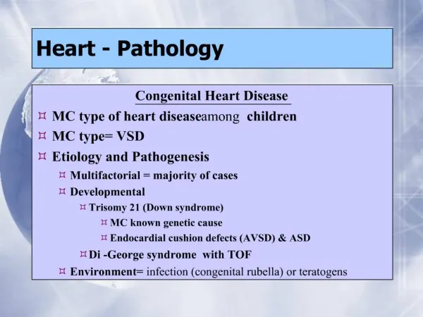
Pathology of the Heart. 4/22/09 Dr. Winokur. The Heart as a Pump. Pump parts - Cardiac equivalent Powersource - Blood supply/oxygen Motor - Myocardium Pump with valves - Cardiac valves Control circuit - Conducting system and neurohumoral control.

E N D
Pathology of the Heart 4/22/09 Dr. Winokur
The Heart as a Pump • Pump parts - Cardiac equivalent • Powersource - Blood supply/oxygen • Motor - Myocardium • Pump with valves - Cardiac valves • Control circuit - Conducting system and neurohumoral control
Pathologic consequences of pump failure • Blood vessels/Oxygen supply - Ischemic heart disease • Myocardium - Cardiomyopathy • Valves - Inadequate forward flow/Increased back pressure • Conducting system - Arrythmias • Neurohumoral system - Inadequate compensation for pathologic processes Failure of any of these components can result in inadequate oxygen delivery to peripheral tissues otherwise known as HEART FAILURE
Case 1 • 65 yo man with past medical history significant for diabetes mellitus, hypertension, and hypercholesterolemia presents to the ED complaining of crushing substernal chest pain that radiates to the jaw.
Physical Exam • Tachycardia • Diaphoretic • EKG • Abnormal with Q-wave • Labs • Elevated troponin I and CK-MB
DIAGNOSIS? • Myocardial infarct
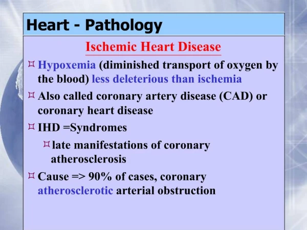
Ischemic heart disease • Mismatch between oxygen supply and demand • Typically results from atherosclerotic narrowing of the coronary arteries • Other causes include vasculitis, intramyocardial coronary arteries • Can result from unusual oxygen demand • Thyrotoxicosis • Tachycardia
Presentation of ischemic heart disease • Chest pain with exertion - Angina pectoris • Myocardial infarct • Many MI s are the initial presentation • Sudden death • Cardiac failure-chronic disease Note that in women the presentation is frequently atypical ie decreased exercise tolerance rather than pain with exercise
Epidemiology • Typically occurs in men in their older than 60 and in women about 10 years later • Risk factors • Hyperlipidemia, especially LDL cholesterol • High blood pressure • Smoking • Diabetes
Coronary pathology of MI • Coronary arteries are partially to completely occluded by atherosclerosis • Significant changes of blood flow occur with >75% narrowing of the arteries • Plaques rupture resulting in thrombosis • Complete occlusion • Myocardial necrosis
Pathology of myocardial infarction • Early changes occur in the 1st .5-1hr and are seen only at the ultrastructural level • Early intervention with thrombolytics or angioplasty can save myocardium • Over the ensuing half day irreversible necrosis occurs and can be identified by the light microscope • By 24 hours there is clear necrosis and neutrophils begin to invade the infarct • This is the first time the infarct is identifiable by gross examination.
Serum enzymes in MI 72 hrs 24 hrs 48 hrs Troponin CK-MB
Over the ensuing week the infarct is overrun by neutrophils, then the dead tissue is removed by macrophages • The infarct is repaired by granulation tissue followed by fibrosis
Complications of myocardial infarction • Cardiac arrhythmias • Cardiac failure/cardiogenic shock • Extension of infarct • Thromboembolism • Ventricular rupture • Papillary muscle rupture • Ventricular aneurysm • Post MI pericarditis
Other presentations of ischemic heart disease • Sudden death • Frequently cause cannot be determined but there is a strong association with coronary atherosclerosis • Chronic ischemia with heart failure • Poor oxygenation results in myocardial atrophy and some myocyte loss resulting in poor cardiac performance • Revascularization can help in large vessel disease
Myocarditis/Myocardial Inflammation • Can be caused by infections • Viral • Bacterial • Fungal • May be secondary to infections resulting in an autoimmune inflammation of the myocardium • Post viral • Post bacterial- Rheumatic heart disease • Autoimmune diseases-Lupus
Motor failure/cardiomyopathy • The heart muscle can fail from primary or secondary causes • Primary dysfunction is related to genetic diseases • Secondary cardiomyopathies result from toxic, infectious and degenerative diseases
Three types of cardiomyopathy • Cardiomyopathy is a primary disease of the heart muscle (excludes myocardial changes resulting from hypertension, valvular disease, ischemic disease and pericardial disease) • Dilated • The ventricular chamber is dilated and the myocardium is modestly thickened • Hypertrophic • The myocardium is markedly thickened especially the septum • Restrictive • The myocardium is can be of normal thickness but it is stiff and unable to relax in diastole
Dilated cardiomyopathy • Causes include genetic, viral/autoimmune and toxic insults • Many cases are idiopathic and are thought to be secondary to previous viral infections • Alcohol is the most common toxic cause • Patients frequently present in heart failure with huge hearts and poor contractility • The prognosis of this condition is poor • 5 year survival is <50% • Patients die from heart failure and arrhythmias
Hypertrophic cardiomyopathy • Primarily a genetic disease and may persist subclinically • Patients present with dyspnea, syncope or sometimes with sudden death • Echocardiography is the best diagnostic modality but may be detected on ECG and physical exam • Pathology- marked hypertrophy of the left ventricle with septal thickening • Septal hypertrophy causes outflow tract obstruction • These patients can frequently be successfully managed
Restrictive cardiomyopathy • Fibrosis or infiltration of the myocardium causes marked stiffness and poor relaxation • Causes include fibrosis, amyloid deposition, sarcoidosis, hemochromatosis, storage diseases • Cardiac filling is impaired and patients present with diastolic heart failure • Poor prognosis unless the underlying cause can be treated
Cardiac hypertrophy • There are two patterns of hypertrophy • Concentric hypertrophy • Caused by pressure overload ie hypertension, valvular stenosis • Results in marked wall thickening with a smaller chamber • Good contractility but poor relaxation • Eccentric hypertrophy • Caused by volume overload ie valve regurgitation or septal defects • Results in wall thickening with dilation of the chamber • Good contractility and acceptable relaxation
Cell length New sarcomeres added lengthwise
Cor Pulmonale • Right sided hypertrophy secondary to pulmonary hypertension followed by dilatation and right heart failure • Acute - 2° to pulmonary thromboembolism • Chronic - Secondary to primary pulmonary hypertension or chronic obstructive pulmonary disease (COPD)
Valvular disease • All of the four valves are subject to disease • Left sided valves are more commonly affected and produce more problems • Diseases include degenerative, infectious and autoimmune
Infectious endocarditis • Damage to the valve surface provides a site for bacterial adherence • Chronic valve disease • Prosthetic valves • Bacteria in the blood stream adhere to the surface and proliferate • Bacteria can be derived from oral cavity, other bacterial infections or the GI tract during procedures • Bacteria can be injected by IV drug abusers and result in right sided endocarditis
Infection can be indolent growth of bacterial colonies or highly destructive infection with valve destruction and incompetence • Strep viridans typically results in an indolent infection • Staph aureus is highly destructive • Prosthetic valves are frequently infected by coagulase negative Staph species • Aggressive and indolent bacteria can embolize and produce peripheral abcesses including the CNS
Non bacterial thrombotic endocarditis (NBTE) • Small vegetations usually occuring at the valve closure lines • Associated with other diseases especially adenocarcinomas and cachexia • Usually asymptomatic and discovered incidentally • Can undergo bacterial colonization leading to infectious endocarditis
Rheumatic fever • An acute, immunologically mediated, multisystem inflammatory disease that follows an untreated episode of group A streptococcal pharyngitis after an interval of a few weeks • Relatively rare in developed countries • Peak incidence is 5-15 yo. • Inflammatory infiltrates may occur in a wide range of sites including the heart
Acute Rheumatic Carditis • Inflammatory changes in all three layers of the heart • Pericardium – fibrinous pericarditis; effusions • Myocardium – heart failure • Endocardium – valvular damage
Rheumatic endocarditis • Repeated episodes of damage eventually damage the valve and associated apparatus • Results in valve stenosis with or without regurgitation • Mitral and aortic valves are most affected • 99% of mitral stenosis is secondary to rhd • Virtually the only cause of simultaneous mitral and aortic stenosis • Can be the substrate for infectious endocarditis
Calcific aortic stenosis • Most common cause of aortic stenosis • Irregular calcium deposits behind valve cusps • Congenitally bicuspid valves • Normal valves as an age-related degenerative change
Mitral valve prolapse • Most frequent valvular lesion (7%) • Young women • Stretching of posterior mitral valve leaflet • Systolic murmur with midsystolic click • Can result in mitral insufficiency • Predisposes to infective endocarditis