Download
1 / 20
210 likes | 255 Views
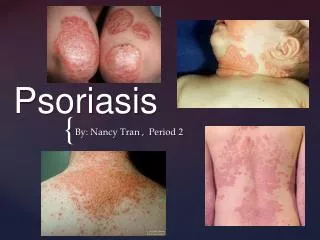
Psoriasis. Background. Psoriasis is an inflammatory, noncontagious, genetically determined skin disorder that most commonly appears as inflamed, edematous skin lesions covered with a silvery white scale. Up to 2% of the population develop Ps during their lifetime.

E N D
Background • Psoriasis is an inflammatory, noncontagious, genetically determined skin disorder that most commonly appears as inflamed, edematous skin lesions covered with a silvery white scale. • Up to 2% of the population develop Ps during their lifetime. • Stress, trauma and infections may induce Ps in susceptible individuals.
Risk Factors • Ps can be present at any time from the first few weeks of life until 80 or more years of age • Most patients experience onset in the third decade of life • There is a definite familial tendency to inherit Ps: when one parent is affected, there is a one in four chance of Ps in each child; with two affected parents, the rate is one in two • Some genetic linkage between Ps and the HLA-Cw6 phenotype
Pathophysiology • The pathological process is a combination of epidermal hyperproliferation and activation of inflammatory pathways with accumulation of inflammatory cells • The hyperplasia of the epidermis results from both a shortened epidermal cell cycle (from an average normal turnover of 23 days to 3-5 days) and an increase in the proliferative cell population • Autoimmune function • Significant evidence is accumulating that psoriasis is an autoimmune disease. • Lesions of psoriasis are associated with increased activity of T cells in underlying skin. • Guttate psoriasis has been recognized to appear following certain immunologically active events, such as streptococcal pharyngitis, cessation of steroid therapy, and use of antimalaria drugs. • Superantigens and T cells • Psoriasis is related to excess T-cell activity. Experimental models can be induced by stimulation with streptococcal superantigen, which cross-reacts with dermal collagen. This small peptide has been shown to cause increased activity among T cells in patients with psoriasis but not in control groups. • Also of significance is that 2.5% of those with HIV develop psoriasis during the course of the disease
Pathophysiology - Details • An infiltrate comprising activated T cells is localized in the dermal papillae of skin and the sublining layer of the joint synovium. Other key cells include various forms of dendritic cells and macrophages; B cells are present but their role is as yet not well defined. These cells generate a number of proinflammatory cytokines, such as tumor necrosis factor (TNF)-alpha, interleukin (IL)-1, and IL-6, which in turn contribute to activation of additional pathogenic cells. In the skin, through the influence of proinflammatory cytokines such as TNF-alpha, keratinocytes proliferate and have prolonged survival, leading to skin thickening and plaque. Proinflammatory cytokines such as TNF-alpha are instrumental in activating endothelial cells to express adhesion molecules such as intercellular adhesion molecule-1 (ICAM-1), vascular adhesion molecule-1 (VCAM-1), and E-selectin (upregulated more in the skin), which promote lymphocyte migration to sites of inflammation. Cutaneous lymphocyte-associated antigen (CLA) is expressed on lymphocytes homing to skin lesions, but not to areas of synovitis. • Vascular pathology is similar in both the skin and synovium, characterized by prominent angiogenesis with abnormal tortuous vessels. Upregulation of angiogenic growth factors, partly via the action of TNF-alpha, including vascular endothelial growth factor (VEGF), transforming growth factor beta (TGF-beta), platelet-derived growth factor (PDGF), and angiopoietins contribute to this phenomenon. Neovascularization is an important component of the inflammatory, erosive nature of the disease. TNF-alpha also contributes to the increased production of proteases such as matrix metalloproteinases (MMPs), which are involved in cartilage destruction. • Abnormal bone remodeling is a key feature of Ps arthritis. Joint-space narrowing and joint erosions occur, as well as osteolysis of both digital tufts and joint areas. However, juxta-articular periosteal new bone formation occurs, as well as ankylosis.
Histopathology – Cardinal Features • Marked thickening of the epidermis (acanthosis) • Absence of the granular layer • Retention of the nuclei in the horny layer (parakeratosis) • Accumulations of polymorphs in the horny layer (Munro’s microabscesses) • Dilated capillary loops in the upper dermis.
Clinical Patterns of Psoriasis • Classical plaque (psoriasis vulgaris) • Scalp Ps • Nail Ps • Guttate • Flexural • “Brittle” • Erythrodermic • Acute pustular (of von Zumbusch) • Chronic palmoplantar pustular (of Barber) • Arthropathic Ps
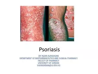
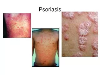
Classical Plaque Psoriasis (psoriasis vulgaris) • The commonest pattern • Single or multiple red plaques (papules) varying from a few millimeters to several centimeters in diameter with a scaly surface • Psoriatic step-by-step triad obtained by scraping: • silvery (stearin) staining • terminal (wet) plate • cappilary-point haemorrhage (Auspitz sign) • Predilection for extensor surfaces: the knees, the elbows and the base of the spine • Lesions are often symmetrical • The scalp and nails are often affected and the arthropathy may also occur • Psoriatic plaques may appear at the site of trauma or scarring – Koebner or isomorphic phenomenon
Scalp Psoriasis • Scalp may be affected alone • Can be difficult to distinguish from severe seborrhoeic dermatitis • Lesions vary from one or two plaques to a sheet of thick scale covering the whole scalp surface • Often, very thick plaques develop, especially at the occiput (nape) • Even decades of persistent scalp Ps have remarkable little effect on the hair, but hair loss is not as uncommon as previously stated.
Nail Psoriasis • Nail abnormalities are frequent and are almost always present in arthropathic Ps • Two common findings can occur together or alone: pitting and onycholysis. • Psoriatic nail pits are relatively large and irregularly arranged • Onycholysis (lifting of the nail plate) initially produces a dull red area with a salmon-pink rim, but the nail becomes brown or yellow in time.
Guttate Psoriasis • Often develops suddenly and may follow an infection, especially a streptococcal sore throat • It is common in adolescents and young adults • Lesions are about one centimeter in diameter and are usually round in shape • Itch is common • Lesions can enlarge and become plaque Ps
Flexural Psoriasis (inverse psoriasis) • Lesions may occur in the groin, natal cleft, axillae, umbilicus, submammary and gluteal folds • Psoriatic balanitis is a form of inverse Ps, that is represented by erythematous plaques on the glans penis • Maceration inevitably occurs, and the scale surface is often lost, leaving a beefy erythematous appearance • It is often itchy
Brittle Psoriasis (instable Ps) • Lesions consist of thin, irritable scaly areas • Lesions may arise de novo or develop suddenly in a patient whose Ps has been stable for years • Systemic steroid therapy and potent topical steroids can induce stable Ps to become “brittle”\ • Lesions may rapidly generalize, leading to erythroderma or acute pustular Ps
Erythrodermic Psoriasis • When psoriatic plaques merge to involve most, or all, of the skin a state of erythroderma or exfoliative dermatitis results; it may appear de novo • The skin is red, hot and scaly; hair and nail loss can develop; itching is severe • There may be a generalized lymphadenopathy • There is a loss of control of temperature regulation accompanied by bouts of shivering • Complications: cardiac failure, renal failure, sudden death due to central hypothermia.
Acute Pustular Psoriasis (of von Zumbusch) • This is a life-threatening condition • Patients with or without pre-existing Ps suddenly develop widespread erythema, superimposed on which are pustules • Pustules can coalesce into lakes of pus (Kogoj-Lapierre pustules) • The pustules are sterile • The patient has a high, swinging fever and is toxic and unwell, with a leucocytosis • If untreated, may die, often of secondary infections.
Chronic Palmo-Plantar Psoriasis (of Barber) • It is unusual for patients to have chronic palmo-plantar pustulosis in association with other forms of Ps • The typical changes consist of erythematous patches with numerous pustules • These gradually change into brown, scaly spots and peel off • Lesions may involve a small area of one hand or foot, or cover the entire surface of both palms and soles • This may lead to considerable disability
Arthropathic Psoriasis • One of the most unpleasant complication of Ps is arthropathy, affecting up to 10% of psoriatics • There are four basic clinical patterns: • distal interphalangeal joint involvement (DIP form) • seronegative rheumatoid-like joint changes • large joint mono- or polyathropathy • spondylitis • Psoriatic arthropathy is erosive and may result in joint destruction • Psoriatics who develop the spondylitic for are usually HLA B27 positive, as in Reiter’s syndrome.
Treatment of Psoriasis • It is an old adage that if there are many treatments for a disease, none works perfectly – this is certainly true of psoriasis • Although each modality is useful in some patients, all represent a compromise in terms of safety, effectiveness and convenience • Many patients require a regimen of different agents for different sites at different times.
Treating Psoriasis Topical Agents • Emollients • Tar • Salicylic acid • Topical steroids • Dithranol (anthralin) • Vitamin D analogues (calcipotriol, tacalcitol, etc) • Vitamin A analogues (tretinoin, tazaroten, etc.) • Ultraviolet radiation (UVA, UVB)
Treating PsoriasisSystemic Agents • PUVA (psoralen + ultraviolet A) • Retinoids (acitretin, etretinate, isotretinoin) • Cytotoxics (methotrexate, azathioprine, hydroxiurea) • Systemic steroids • Ciclosporin • TNFα inhibitors (infliximab, etanarcept, etc.)