Download
1 / 37
390 likes | 553 Views
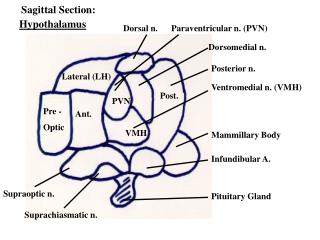
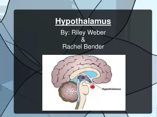
Hypothalamus. Historical Perspective. Wilhelm His (1893) Distinct division of diencephalon Harvey Cushing (1920s), neurosurgeon Diabetes insipidus: excess water excretion & thirst Cushing syndrome: excess secretion of cortisol 1930s: cytoarchitectural definition of nuclei

E N D
Historical Perspective • Wilhelm His (1893) • Distinct division of diencephalon • Harvey Cushing (1920s), neurosurgeon • Diabetes insipidus: excess water excretion & thirst • Cushing syndrome: excess secretion of cortisol • 1930s: cytoarchitectural definition of nuclei • Functional specialization (Ranson, Hess) • Lesion, electrical stimulation, affect appetite, body weight, water balance, autonomic control, reproductive function, emotional behavior
Historical Perspective • Ernst & Berta Scharrer (1940): first evidence of neurosecretary neurons (neurons in the brain that secrete hormones into blood stream) • Wolfgang Bargmann (1949): secretory neurons, cell bodies in hypothalamus & axons in posterior pituitary • Harris & Green (1950s): neurohumoral regulation of anterior pituitary; identified neurovascular link, portal plexus • Guillemin & Schally (1970s): characterized peptide hormones that act on anterior pituitary
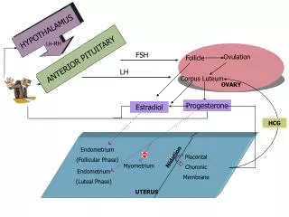
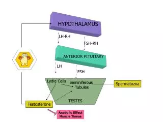
Cushing Syndrome Adrenal cortex secretes too much cortisol. Results from tumor in pituitary or adrenal cortex. Cortisol causes proteins to break down. Muscle atrophy, skin thinner, bones stop growing & easily fractured, lymphatic tissue shrinks -reduced resistance to infection, fat redistributed from arm & legs to face & trunk
Olfactory input • Multi-synaptic pathways • olfactory tubercle, piriform cortex, amygdala • Medial forebrain bundle • Stria terminalis • Ventral amygdalofugal pathway • Fornix • Reproduction, defense, feeding
Visual input • Impose temporal organization • SCN • Central visual projection • Direct projection from ganglion neurons of retina • SCN outputs sparse, confined largely to hypothalamic structures
Visceral input • Nucleus of the Solitary tract (NST), or nucleus tractus solitarius (NTS) • Principal visceral sensory nucleus, input from major organs via VII, IX, X • Taste, GI, cardiovascular, respiratory • Direct: to PVN, LH • Indirect: via ventralateral medulla & parabrachial nucleus of pons
Multi-modal brainstem afferents • Medial forebrain bundle (bidirectional); major conduit • Monoamine containing neurons • Locus Coeruleus & lateral tegmental cell groups (NE) • Brainstem raphe (5-HT) • Dopaminergic neurons • Substantia nigra & ventral tegmental area • Former - movement; latter - motivation, addiction
Limbic inputs • Hippocampus via fornix • Subicular complex to mamillary bodies (postcomissural fornix); direct route • Hippocampus proper (Ammon’s horn) via septum to all longitudinal levels of hypothalamus • Amygdala • Stria terminalis • Vental amygdalofugal pathway
Non-synaptic input • Circumventricular organs • Subfornical organ, SFO; vascular organ of the lamina terminalis, OVLT; median eminence, ME; posterior pituitary; pineal gland; subcommissural organ, SCO; area postrema, AP. • OVLT, ME, PP in hypothalamus • SFO & AP have extensive connections with hypothalamus