Download
1 / 30
300 likes | 452 Views
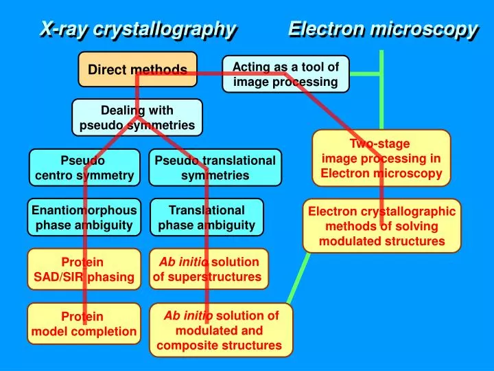
X-ray crystallography. Electron microscopy. Direct methods. Acting as a tool of image processing. Dealing with pseudo symmetries. Two-stage image processing in Electron microscopy. Pseudo centro symmetry. Pseudo translational symmetries. Enantiomorphous phase ambiguity.

E N D
X-ray crystallography Electron microscopy Direct methods Acting as a tool of image processing Dealing with pseudo symmetries Two-stage image processing in Electron microscopy Pseudo centro symmetry Pseudo translational symmetries Enantiomorphous phase ambiguity Translational phase ambiguity Electron crystallographic methods of solving modulated structures Protein SAD/SIR phasing Ab initio solution of superstructures Protein model completion Ab initio solution of modulated and composite structures
电子显微学 X - 射线晶体学 直接法 用于图像处理 处理赝对称性 两步法电子 显微学图像处理 赝中心对称 赝平移对称 电子晶体学方法 求解调制结构 蛋白质SAD/SIR 相位推演 超结构 从头求解 蛋白质 模型完整化 从头测定调制结构 和组合结构
Modulated and Composite structures Software Initial phasing and Model completion of Proteins Image processing in Electron Microscopy
Enantiomorphous phase ambiguity resolved by the direct method Enantiomorphous phase ambiguity resolved by the direct method Sample: ZCW; Space group: P21 Acta Phys. Sin.27, 169-174 (1978) K Before treatment After treatment Direct method result Final map c c a c b b
X-ray Crystallography Diffraction wave front Object Patterson map Diffraction pattern Fourier transform Fourier transform Electron microscopy Objective lens Image plane Back focal plane Object Diffraction pattern Blurred image
Objective lens Image plane Back focal plane Object Multiplied with a contrast transfer function Diffraction pattern Blurred image X-ray Crystallography Diffraction wave front Object Structure image blurred by convoluting with It’s inverse Multiplied with F*h Patterson map Diffraction pattern Fourier transform Fourier transform Electron microscopy Structure image blurred by convoluting with the Fourier transform of a contrast transfer function
X-ray Crystallography Diffraction wave front Object Multiplied with F*h Patterson map Diffraction pattern Objective lens Image plane Back focal plane Object Multiplied with a contrast transfer function Diffraction pattern Blurred image In the derivation of X-ray diffraction phases, direct methods are acting as a tool of image processing. They convert the blurred image - Patterson map - to the deblurred image - electron density map. It is reasonable to expect that direct methods can be used in high-resolu-tion electron micro-scopy as a tool of image processing. Fourier transform Fourier transform Electron microscopy
B Phase ambiguity intrinsic in SAD F-* F+ <F> Fo F” |Dj | |Dj | j” j - F’ j’ j + A - F”
B Phase ambiguity intrinsic in SIR FN |DjN| |DjN| FD jR jIIN jIN FR |DjD| jIID |DjD| jID jR A
D – derivative, R – replacing atoms, N – native • – SIR phase doublet
SAD/SIR data P+(Djh) = 1/2 Eh ,j’h,|Djh| Calculate mhandDjh,best End Calculate P+(Djh)
Lysozyme Space group: P43212 Unit cell: a = 78.4, c = 37.0Å Number of residues in the ASU: 129 Resolution limit: 2.01Å; Multiplicity: 47.2 Anomalous scatterer: S (10 ) X-rays: Cr-Ka, l = 2.291Å, Df ” = 1.14 Bijvoet ratio: <|DF |>/<F > = 2.4% Data provided by Prof. Isao Tanaka and Dr. Nobuhisa Watanabe
Azurin Space group: P4122 Unit cell: a = b = 52.65, c = 100.63Å Number of residues in the ASU: 129 Resolution limit: 1.9Å Multiplicity: 10.0 Anomalous scatterer: Cu (1) X-rays: synchrotron radiation l = 0.97ÅDf” = 2.206 Bijvoet ratio: <|DF|>/<F> = 1.44% Data provided by Prof. S. Hasnain
Xylanase Space group: P21 Unit cell: a = 41.07, b = 67.14, c = 50.81Å b = 113.5o Number of residues in the ASU: 303 Resolution limit: 1.75Å; Multiplicity: 15.9 Anomalous scatterer: S(5 ) X-rays:synchrotron radiation l = 1.488Å; D f ” = 0.52 Bijvoet ratio:<|DF |>/<F > = 0.56% Data courtesy of Dr. Z. Dauter, National Cancer Institute, USA
Alanine racemase Space group: C 2 2 21 Unit cell: a = 72.68 b = 76.13, c = 136.27Å Number of residues in the ASU: 357 Resolution limit: 2.3Å Data collection range: 360o Anomalous scatterer: Se (8), S (6), P (1) X-rays: Cr-Ka, l = 2.291Å Bijvoet ratio: <|DF |>/<F > = 2.8% Data provided by Dr. Cheng Yang
Alanine racemase Space group: C 2 2 21 Unit cell: a = 72.68 b = 76.13, c = 136.27Å Number of residues in the ASU: 357 Resolution limit: 2.3Å Data collection range: 360o Anomalous scatterer: Se (8), S (6), P (1) X-rays: Cr-Ka, l = 2.291Å Bijvoet ratio: <|DF |>/<F > = 2.8% Data provided by Dr. Cheng Yang
SOLVE/RESOLVE + OASIS/DM SIR SOLVE/RESOLVE
SOLVE/RESOLVE + OASIS/DM SOLVE/RESOLVE SAD
SOLVE/RESOLVE + OASIS/DM SIR SAD SIR SOLVE/RESOLVE + OASIS/DM SOLVE/RESOLVE SOLVE/RESOLVE
SAD SOLVE/RESOLVE + OASIS/DM SOLVE/RESOLVE + OASIS/DM SOLVE/RESOLVE SOLVE/RESOLVE
2.1Å 3.0Å 4.0Å 3.5Å