Download
1 / 1

E N D
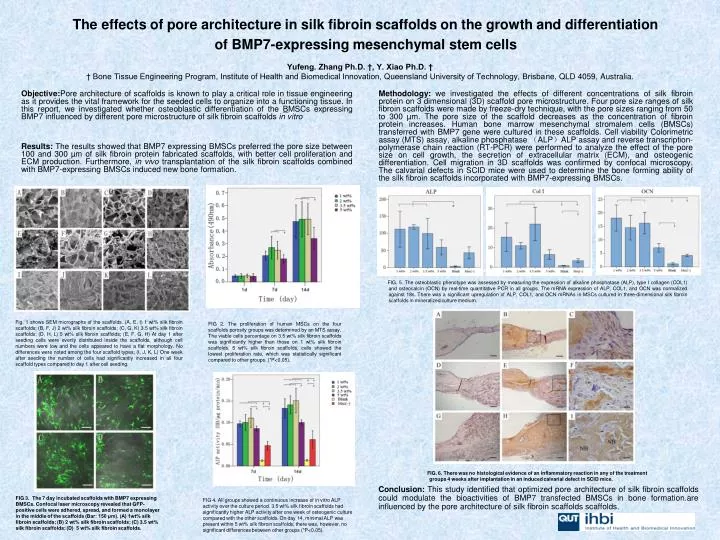
Objective:Pore architecture of scaffolds is known to play a critical role in tissue engineering as it provides the vital framework for the seeded cells to organize into a functioning tissue. In this report, we investigated whether osteoblastic differentiation of the BMSCs expressing BMP7 influenced by different pore microstructure of silk fibroin scaffolds in vitro Methodology:we investigated the effects of different concentrations of silk fibroin protein on 3 dimensional (3D) scaffold pore microstructure. Four pore size ranges of silk fibroin scaffolds were made by freeze-dry technique, with the pore sizes ranging from 50 to 300 µm. The pore size of the scaffold decreases as the concentration of fibroin protein increases. Human bone marrow mesenchymal stromalem cells (BMSCs) transferred with BMP7 gene were cultured in these scaffolds. Cell viability Colorimetric assay (MTS) assay, alkaline phosphatase (ALP)ALP assay and reverse transcription-polymerase chain reaction (RT-PCR) were performed to analyze the effect of the pore size on cell growth, the secretion of extracellular matrix (ECM), and osteogenic differentiation. Cell migration in 3D scaffolds was confirmed by confocal microscopy. The calvarial defects in SCID mice were used to determine the bone forming ability of the silk fibroin scaffolds incorporated with BMP7-expressing BMSCs. The effects of pore architecture in silk fibroin scaffolds on the growth and differentiation of BMP7-expressing mesenchymal stem cells Results:The results showed that BMP7 expressing BMSCs preferred the pore size between 100 and 300 µm of silk fibroin protein fabricated scaffolds, with better cell proliferation and ECM production. Furthermore, in vivo transplantation of the silk fibroin scaffolds combined with BMP7-expressing BMSCs induced new bone formation. FIG. 5. The osteoblastic phenotype was assessed by measuring the expression of alkaline phosphatase (ALP), type I collagen (COL1) and osteocalcin (OCN) by real-time quantitative PCR in all groups. The mRNA expression of ALP, COL1, and OCN was normalized against 18s. There was a significant upregulation of ALP, COL1, and OCN mRNAs in MSCs cultured in three-dimensional silk fibroin scaffolds in mineralized culture medium. Yufeng. Zhang Ph.D. †, Y. Xiao Ph.D. † † Bone Tissue Engineering Program, Institute of Health and Biomedical Innovation, Queensland University of Technology, Brisbane, QLD 4059, Australia. Fig. 1 shows SEM micrographs of the scaffolds. (A, E, I) 1 wt% silk fibroin scaffolds; (B, F, J) 2 wt% silk fibroin scaffolds; (C, G, K) 3.5 wt% silk fibroin scaffolds; (D, H, L) 5 wt% silk fibroin scaffolds; (E, F, G, H) At day 1 after seeding cells were evenly distributed inside the scaffolds, although cell numbers were low and the cells appeared to have a flat morphology. No differences were noted among the four scaffold types; (I, J, K, L) One week after seeding the number of cells had significantly increased in all four scaffold types compared to day 1 after cell seeding. FIG 2. The proliferation of human MSCs on the four scaffolds porosity groups was determined by an MTS assay. The viable cells percentage on 3.5 wt% silk fibroin scaffolds was significantly higher than those on 1 wt% silk fibroin scaffolds. 5 wt% silk fibroin scaffolds, cells showed the lowest proliferation rate, which was statistically significant compared to other groups. (*P<0.05). FIG. 6. There was no histological evidence of an inflammatory reaction in any of the treatment groups 4 weeks after implantation in an induced calvarial defect in SCID mice. Conclusion:This study identified that optimized pore architecture of silk fibroin scaffolds could modulate the bioactivities of BMP7 transfected BMSCs in bone formation.are influenced by the pore architecture of silk fibroin scaffolds scaffolds. FIG 3. The 7 day incubated scaffolds with BMP7 expressing BMSCs. Confocal laser microscopy revealed that GFP-positive cells were adhered, spread, and formed a monolayer in the middle of the scaffolds (Bar: 150 μm). (A) 1wt% silk fibroin scaffolds; (B) 2 wt% silk fibroin scaffolds; (C) 3.5 wt% silk fibroin scaffolds; (D) 5 wt% silk fibroin scaffolds. FIG 4. All groups showed a continuous increase of in vitro ALP activity over the culture period. 3.5 wt% silk fibroin scaffolds had significantly higher ALP activity after one week of osteogenic culture compared with the other scaffolds. On day 14, minimal ALP was present within 5 wt% silk fibroin scaffolds; there was, however, no significant differences between other groups (*P<0.05).