Download
1 / 27
281 likes | 680 Views
General Approach in Investigation of Haemostasis. Lecture 1: Introduction. Preanalytical Variables including. Sample Collection. Site Selection. Storage Requirements. Transportation of Specimen. Haemostasis.

E N D
General Approach in Investigation of Haemostasis Lecture 1: Introduction
Preanalytical Variables including Sample Collection. Site Selection. Storage Requirements. Transportation of Specimen.
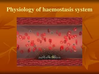
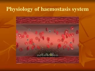
Haemostasis Hemostasis is a complex interaction between vessels, platelets and coagulation proteins that, when working properly, stops bleeding while maintaining blood flow in the vessel. Specific tests are available to evaluate platelet function, coagulation proteins, natural occurring inhibitors and fibrinolysis.
Sample Collection Proper sample collection is of utmost importance for reliable test results to evaluate the bleeding patient, thrombosis or fibrinolysis (preanalytical phase) All these tests are influenced by sample collection, sample processing and sample storage. The laboratory will not evaluate samples that are hemolyzed, clotted, contain fibrin strands or improperly stored. Reference Laboratory Services will immediately notify the client of any problems with the sample. When blood is withdrawn from a vessel, changes begin to take place in the components of blood coagulation. Some occur almost immediately, such as platelet activation and the initiation of the clotting mechanism dependent on surface contact.
Sample Collection * CLSI : Clinical and Laboratory Standards Institute • Anticoagulant of choice • 3.8% or 3.2% Sodium Citrate • 3.2 % Preferred as the standard measure due to stability and closeness to the plasma osmolality • Anticoagulant/blood ratio is critical (1:9) • Exact amount of blood must be drawn. No short draws are acceptable, this will falsely increase results due to presence of too much anticoagulant • CLSI guideline is +/- 10 % of fill line • Purpose of the anticoagulant is to bind or chelate calcium to prevent clotting of specimen
Sample Collection • Other anticoagulants, including oxalate, heparin, and EDTA, are unacceptable. • The labile factors (factors V and VIII) are unstable in oxalate, whereas heparin and EDTA directly inhibit the coagulation process and interfere with end-point determinations. • Additional benefits of trisodium citrate are that the calcium ion is neutralized more rapidly in citrate, and APTT tests are more sensitive to the presence of heparin.
Sample Collection : Samples with High hematocrits * National Committee for Clinical Laboratory Standards • According to the latest CLSI (formerly NCCLS) guideline on coagulation testing, it is important to adjust the sodium citrate volume when a patient’s hematocrit is greater than 55%. • Examples of patients who may have elevated hematocrit values are newborns or people with polycythemia vera. • NCCLS* recommends adjusting anticoagulant ratio for patients with hematocrits exceeding 55% • High hematocrits may cause falsely prolonged test results due to an over- anticoagulated sample • Formula correction achieves a 40% hematocrit
X = (100–PCV)*vol./(595–PCV) Where: X= volume of sodium citrate Vol =volume of whole blood drawn PCV= patient’s hematocrit Examples: Patients Hct= 60%, V= 5 mL X=(100-60)*5 / (595-60) = 40*5 / 535 = 0.34 ml Patient Hct = 25%, V=5 ml X=(100-25)*5 / (595-25) = 75*5 / 570 = 0.65 ml
Site Selection • Untraumaticvenipuncture is required • Traumatic venipunctures release tissue factor and initiate coagulation • Fingersticks/Heelsticks are not allowed • Indwelling IV line draws are discouraged • Contain heparin & diluted blood • Falsely increased results • Order of Draw • Evacuated tube system • Blue top is 2nd • If 2nd tube drawn, 1st top must be anticoagulant free (i.e. red top)
Storage Requirements • Prothrombin Time: PT • Uncentrifuged or centrifuged with plasma remaining on top of cells in unopened tube kept at 2-4 oC or 18-24 oC must be tested within 24 hours of collection • Activated Partial Thrombin Time: APTT • Uncentrifuged or centrifuged with plasma remaining on top of cells in unopened tube kept at 2-4 oC or 18-24 oC must be tested within 4 hours of collection • Other Assays • Fibrinogen, Thrombin Time, Factor Assays • Centrifuged with plasma remaining on top of cells in unopened tube kept at 2-4 oC or 18-24 oC must be tested within 4 hours of collection
StorageRequirements • Other general notes • Perform coagulation tests ASAP • Specimen may deteriorate rapidly (especially factors V and VIII) • If the testing is not completed within specified times, plasma should be removed from the cells and placed in a frost free freezer • - 20 oC for two weeks • -70 oC for six months
Transportation of Specimen Send specimen on ice OR deliver to lab ASAP Separate cells from plasma immediately via centrifugation
Platelet Poor Plasma • Platelet –Poor plasma (PPP) • Platelet-Poor plasma is necessary for coagulation testing to prevent activation of platelets and release of PF4, a heparin inhibitor. • The plasma platelet count must be < 10,000 /mm3. • Specimen has been centrifuged for 15 minutes @ 2500 x g • Why is PPP essential? • Contains platelet factor 4 (heparin neutralizer) • Contains phospholipids (affects lupus anticoagulant and factor assay testing) • Contains proteases (affect testing for vWF)
Platelets Poor Plasma preparation: To prepare platelet-Poor plasma Centrifuge the blue top evacuated tubes (CLSI, formerly NCCLS recommendation is 1500 rpm for 15 minutes). Using a plastic pipette, immediately remove the top 2/3 of the plasma to a plastic aliquot tube. Centrifuge this plasma sample and remove the top ¾ of the plasma to a second plastic aliquot tube with a fresh plastic pipette. Freeze the specimen within one hour of collection.
Platelets Rich Plasma (PRP) • Platelet-Rich plasma (PRP) • Used in platelet function studies • 200-300 x 10 9 /L • Specimen must be centrifuged for 10 minutes @ 200 x g
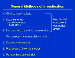
Principles of Laboratory Analysis The more detailed investigations of coagulation proteins also require caution in their interpretation depending on the type of assay performed. These can be divided into three principal categories, as described in the following sections. Immunological Assays Using Chromogenic Peptide Substrates (Amidolytic Assays) Coagulation Assays Other Assays
Immunological Include immuno-diffusion, immuno-electrophoresis, radioimmunometric assays, latex agglutination tests, and tests using enzyme-linked immunosorbent assays (ELISA). Fundamentally, all these tests rely on the recognition of the protein in question by polyclonal or monoclonal antibodies. Polyclonal antibodies lack specificity but provide relatively high sensitivity, whereas monoclonal antibodies are highly specific but produce relatively low levels of antigen binding.
latex agglutination kit: Latex microparticles are coated with antibodies specific for the antigen to be determined. When the latex suspension is mixed with plasma an antigen–antibody reaction takes place, leading to the agglutination of the latex microparticles. Agglutination leads to an increase in turbidity of the reaction medium, and this increase in turbidity is measured photometrically as an increase in absorbance. Usually the wavelength used for latex assays is 405 nm, although for some assays a wavelength of 540 or 800 nm is used. This type of assay is referred to as immuno- turbidimetric.
Notes: Do not freeze latex particles because this will lead to irreversible clumping. An occasional problem with latex agglutination assays is interference from rheumatoid factor or paraproteins. These may cause agglutination and overestimation of the protein under assay.
Chromogenic Assay Chromogenic, or amidolytic, methodology is based on the use of a specific color-producing substance known as a chromophore. the chromophore normally used in the coagulation laboratory is para-nitroaniline (pNA), which has an optical absorbance peak at 405 nm on a spectrophotometer.
Coagulation Assays Coagulation assays are functional bioassays and rely on comparison with a control or standard preparation with a known level of activity. In the one-stage system optimal amounts of all the clotting factors are present except the one to be determined, which should be as near to nil as possible. The best one-stage system is provided by a substrate plasma obtained either from a patient with severe congenital deficiency or artificially depleted by immuno-adsorption.
Coagulation Assays Coagulation techniques are also used in mixing tests to identify a missing factor in an emergency or to identify and estimate quantitatively an inhibitor or anticoagulant. The advantage of this type of assay is that it most closely approximates the activity in vivo of the factor in question. However, they can be technically more difficult to perform than the other types described earlier.
Other Assays Using snake venoms (The Taipan venom time employs a reagent isolated from the venom of the Taipan snake (Oxyuranus scutellatus) that directly activates prothrombin in the presence of phospholipid and calcium.) Aassay of ristocetin cofactor (used to diagnose von Willebrand disease ) The clot solubility test for factor XIII. DNA analysis is becoming more useful and more prevalent in coagulation. However, this requires entirely different equipment and techniques