Download

1 / 71
810 likes | 1.47k Views
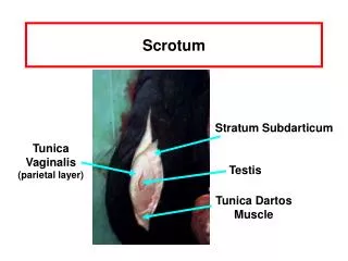
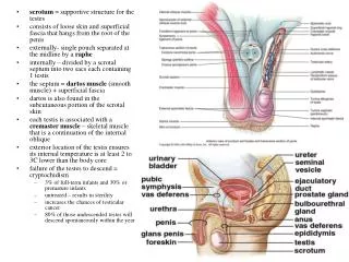
The acute pediatric scrotum. Khosro taheri 89123. Differential dianostic possibilities for the acute scrotum depend the patients age: Neonate: testicular torsion(pre or postnatal), mecunium peritonitis orchitis , hernia and hydrocele

E N D
The acute pediatric scrotum Khosrotaheri 89\12\3
Differential dianostic possibilities for the acute scrotum depend the patients age: Neonate:testicular torsion(pre or postnatal),mecunium peritonitis\orchitis , hernia and hydrocele Older childs:testiculartorsion,appendage torsion and epididymitis
Testicular torsion is primary concern. • Although Rapidity of symptom onset,associated symptoms and physical examination findings may suggest the diagnosis, imaging is often required. • Ultrasound with color doppler is the modality of choice.
Testicular torsion : • has two incidence peaks: • one in the perinatal period and • one around puberty. • In the peripubertal period, testicular torsion is intravaginal , with the testis and spermatic cord twisting within the tunica vaginalis caused by imprope fixation of the testis (bell-clapper deformity).
The twisting of the spermatic cord results first in compromised venous outflow and ultimately arterial inflow, leading to testicular ischemia and eventual infarction. Among children and adolescents presenting with acute scrotal pain, spermatic cord torsion is present in 6% to 31%
onset of symptoms is most rapid for testicular torsion. Nausea and vomiting are more common in testicular torsion and have a positive predictive value of more than 96% • Physical examination findings include swelling, an abnormal transverse lie of the testis within the scrotum, and loss of the cremasteric reflex.
If the history and physical examination strongly suggesttorsion, the patient should go directly to surgery without any delay to perform imaging studies imaging should be used for those patients who have unclear diagnoses, in whom torsion is unlikely and another diagnosis needs to be addressed, and in those who have symptoms lasting longer than 24 hours, because even if torsion is present, the chance of testicular salvage is remote and emergency surgery may not be required.
Testicular survival is directly related to the time from onset of symptoms, with only 20% salvage after 12 hours and virtually no salvage after 24 hours.Nonviable testes are removed to prevent immunemediated injury to the contralateral testis. • Early sonographic literature focused on changes in parenchymalechotexture caused by ischemia,which were of limited value. In general, the acutely torsed testis is more hypoechoic than normal. The gray-scale findings in testicular torsion, however, are seldom normal. Useful gray-scale findings for torsion include an abnormal transverse testicular lie and a ratesticular ‘‘mass’’ composed of swollen edematous epididymis and spermatic cord.
In some cases, the actual twist of the spermatic cord can be visualized • Reactive hydrocele and scrotal skin thickening is often present, particularly in later cases.
To diagnose testicular torsion, one must definitively demonstrate reduced or absent blood flow to the symptomatic testis and normal blood flow to the asymptomatic testis. • The sensitivity of color Doppler examination with newer ultrasound equipment in detecting acute testicular torsion in children is 90% to 100%, with the specificity of technically adequate studies being essentially 100%
the hallmark of testicular torsion is absent or diminished arterial flow relative to the asymptomatic contralateral testis • The presence of flow in the painful testis does not exclude the diagnosis of torsion.
. Color Doppler should be used to perform a semiquantitative estimate of blood flow symmetry. Early or partial cord torsion is not uncommon these testes may demonstrate arterial flow, albeit diminished in quantity from the asymptomatic contralateral testis and with diminished diastolic flow
A nonsurgical painful testis must be hyperemic relative to the asymptomatic side, otherwise torsion remains a diagnostic possibility. Exceptions to this are cases of spontaneous detorsion, which results in increased blood flow to the testis and peritesticular tissues, giving the appearance of an inflammatory nonsurgical condition • Spontaneous detorsion is usually accompanied by a marked relief in symptoms, however, which can help clarify the situation.
The sonographic findings in late torsion (greater than 24 hours) become more dramatic, with the testicle often becoming disorganized and heterogeneous because of infarction. Peritesticular soft tissue inflammation becomes marked. Color Doppler shows hyperemia of the scrotum and peritesticular tissues with absent flow to the testis • If not removed, the infarcted testis begins to atrophy, often becoming hyperechoic because of fibrosis or calcification
Extravaginal torsion occurs in neonates because of poor fixation of the spermatic cord within the inguinal canal, these torsions usually occur in utero with the newborn presenting with a firm, discolored scrotum that is usually painless. • more commonly the chronically torsed testis is nearly normal in size and there is a peripheral echogenic rim corresponding to calcifications in the tunica albuginea, indicating a more remote event
Torsion of a testicular or epididymal appendage is the most common cause of acute scrotal pain in children before puberty, accounting for 26% to 67% of patients resenting to pediatric emergency departments • This is probably an underestimate, because appendiceal torsion may account for most cases of prepubertal ‘‘epididymitis,’’because most children do not have any anatomic or infectious predisposition for true epididymitis and the imaging findings can be similar
The appendix testis is present in 92% of males, and the appendix epididymis is present in 25% [12]; thus, the appendix testis is the more common one to torse. • Although appendiceal torsion may mimic testicular torsion, the onset of pain tends to be more gradual than with testicular torsion, and systemic symptoms are usually absent
Appendiceal torsion is not a surgical emergency and responds well to conservative management • Atrophy of the appendage and resolution of symptom with supportive care is the usual outcome. Persistently symptomatic appendages may require surgical removal for pain relief.
On sonography, the torsed appendix appears as a small hyper- or hypoechoic mass adjacent to the superior aspect of the testis or epididymis • Coronal or transverse scanning above the testis may facilitate diagnosis • With careful examination the sensitivity of sonographic detection of appendiceal torsion approaches 90%
Although the torsed appendage has no flow, it incites an intense inflammatory response in the adjacent tissues. Color Doppler imaging demonstrates marked hyperemia focally around the area of torsion or diffusely throughout the entire testis and epididymis
The ultrasound appearance can be dramatic, with severe swelling and edema, scrotal skin thickening, and a reactive hydrocele • Later the torsed appendage may appear as a small hyperechoic or calcified structure adjacent to the testis or may even detach to become an intrascrotal loose body or scrotolith
Acute epididymitis: epididymitis accounts for 28% to 47% of cases of acute scrotal pain among pediatric patients In fact, it is believed that many cases of prepubertalepididymtis (particularly those with negative urine cultures) are actually cases of appendiceal torsion
In younger patients, noninfectious epididymitis can be caused by genitourinary abnormalities, such as an ectopic ureter draining into the vas deferens or seminal vesicles .Physiologic or anatomic bladder outlet obstruction may lead to reflux of urine into • the ejaculatory ducts, producing epididymitis. • Epididymitis can occur after scrotal trauma and it also may be idiopathic without a clear discernible etiology
Imaging reveals epididymal enlargement that may be diffuse or localized to one portion of the epididymis. • The swollen epididymis is usually hypoechoic,but subsequent hemorrhage or edema can produce variable echogenicity. • As with other inflammatory scrotal conditions, there is usually scrotal skin thickening and a reactive hydrocele.
Color Doppler shows increased blood flow in the inflamed epididymis compared with the asymptomatic side • In 20% of patients who have epididymitis, however, the gray-scale appearance of the epididymis is normal and the inflammation is only revealed with color Doppler. • With newer color Doppler technology, the normal epididymis shows minimal detectable flow and the detection of any significant vascularity should be considered abnormal
Orchitis: • In acute epididymo-orchitis,the testis is usually enlarged and diffusely hypoechoic,becoming increasingly heterogeneous overtime as the inflammatory process continues • Focal lesions are less common, but when they occur, they appear as hypoechoic lesions adjacent to areas of epididymal inflammation. Inflammatory foci are often ill defined and can appear mass-like. The clinical scenario, however, usually differentiates infection from tumor.
Color Doppler shows increased blood flow to the inflamed testis, with markedly increased diastolic flow • Similar to epididymitis, gray-scale sonography is normal in 10% to 40% of patients who have orchitis, only being reliably detected with color flow imaging • Acute epididymitis or epididymo-orchitis may produce testicular ischemia if spermatic vessels are compressed by a swollen epididymis or spermatic cord.
Compromise of the draining pampiniform venous plexus and lymphatics is likely more important in producing testicular ischemia than is involvement of the testicular artery • The testis appears enlarged and heterogeneous and has decreased or absent color flow. Pulsed Doppler waveforms demonstrate diastolic flow reversal of arterial waveforms resulting from increased intratesticular impedance caused by decreased venous outflow and vascular congestion
Primary orchitis is less common than secondary epididymo-orchitis and is usually viral in origin. • Testicular inflammation from mumps orchitis occurs in approximately 30% of infected post pubertal boys .Involvement is commonly bilateral, producing enlarged and heterogeneous testes at sonography that are hyperemic with color Doppler • Testicular atrophy may result, and later fertility is probably reduced.
Trauma: • As the physical examination may be limited by pain and swelling, ultrasound plays an important role in evaluating for hematoceles, testicular hematoma, or the more severe testicular fracture or rupture. • Traumatic hematoceles are common and appear acutely as echogenic fluid collections.
as the hematoma organizes and clot retracts, the hematoma takes on the appearance of a complex septated cystic fluid collection • Hematomas confined to the scrotal wall or peritesticular soft tissues also occur, and appear as variably echoic collections depending on their age.
Testicular hematoma results from blunt injury to the scrotum. • As with hematomas elsewhere, the enlarged testis shows areas of increased or decreased parenchymalechogenicity depending on the age of the hematoma. • Acutely, intratesticular hematomas are heterogeneous and may be hypo- or hyperechoic to surrounding testicular tissue
Scrotal wall thickening and hydroceles or hematoceles are associated findings • Color Doppler imaging is vital to prove viability of the traumatized testis and should show a normally perfused testis except for focal areas of absent vascularity in the area of the hematoma • Follow-up imaging may reveal focal or diffuse testicular atrophy resulting from resorption of injured parenchyma or from ischemia from a surrounding hematocele or intratunica swelling
Testicular fracture appears as a hypoechoic band interrupting the testicular parenchyma, but the testicular contour is well defined and the tunica albuginea is intact. • Doppler evaluation is important. If perfusion to all portions of the testis can be identified, conservative management is possible; otherwise, emergent surgical exploration is required
With testicular rupture, the hyperechoic tunica albuginea is interrupted, and there is extrusion of testicular tissue into the scrotum • This can be a difficult sonographicdiagnosis,especially in the presence of a large hematocele The testis appears heterogeneous because of hemorrhage and tissue injury and has ill-defined margins at the site of rupture • Doppler rarely reveals flow, but surgery is indicated for attempted salvage and to remove any nonviable tissue that may lead to abscess formation or impede spermatogenesis in the remaining testis.