Download
1 / 42
440 likes | 745 Views
Exam I. Study Guide. Explain . Parfocal Inversion Summarize the procedure for preparing a wet mount slide. Questions. What is the function of tendons and ligaments? Name the enzyme that breaks down ATP during a muscle contraction. What is the function of red bone marrow?

E N D
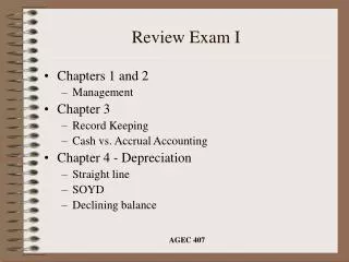
Exam I Study Guide
Explain • Parfocal • Inversion • Summarize the procedure for preparing a wet mount slide
Questions • What is the function of tendons and ligaments? • Name the enzyme that breaks down ATP during a muscle contraction. • What is the function of red bone marrow? • What is the chemical composition of the yellow bone marrow?
Plasma membrane Cytoplasm Organelles Nucleus a) A eukaryotic animal cell has a large nucleus and numerous small organelles. The cytoplasm is enclosed by a flexible plasma membrane.
Question • Identify and label three structures of the animal cells
Question • What appearance will red blood cells have when they are placed in the following solutions? Explain your answer in detail. • Hypertonic • Isotonic • Hypotonic
Figure 5.12 Coxal bones and sacrum (pelvis) Pubic symphysis Femur (upper leg) Patella (knee cap) Tibia Lower leg Fibula 7 Tarsals (ankle) 5 Metatarsals (foot) 14 Phalanges (toe bones)
Spongy bone (spaces contain red bone marrow) Epiphysis Compact bone Yellow bone marrow Blood vessel Diaphysis Periosteum Central cavity (contains yellow bone marrow) Epiphysis a) A partial cut through a long bone. Figure 5.1a
Question • Why do bone injuries heal faster than injuries to cartilage?
Pectoralis major • Draws arm forward and toward the body • Biceps brachii • Bends forearm at elbow • Rectus abdominus • Compresses abdomen • Bends backbone • Compresses chest cavity Figure 6.1a
Deltoid • Raises arm • Trapezius • Lifts shoulder blade • Braces shoulder • Draws head back • Triceps brachi • Straightens forearm at elbow • Latissimus dorsi • Rotates and draws • arm backward and • toward body • Gluteus maximus • Extends thigh • Rotates thigh laterally • Gastrocnemius • Bends lower leg at • knee • Bends foot away from • knee Figure 6.1b
Name and identify the kissing and blinking muscles.
Question • What are the two divisions of the skeletal System? • Name four bones from each division.
Appendicular skeleton Axial skeleton Cranium (skull) Maxilla Mandible Clavicle Scapula Sternum Ribs Humerus Vertebrae Ulna Radius Carpals Metacarpals Sacrum Phalanges Coxal bone Femur Patella Tibia Fibula Tarsals Metatarsals Phalanges Figure 5.5
Osteons • The Osteon: label the osteocytes, lamellea, lacuna and canaliculi. • Why is the central canal larger compared to the other components of bone tissue? • Describe the appearance of the canaliculi and give their function.
Question • Describe how an osteocyte located near a central canal can pass nutrients to osteocytes located far from the central canal.
Identify the following structures • Sarcomere • Motor neuron • Sarcoplasmic reticulum • T-tubules • Actin and myosin
True ribs= 7False ribs= efloating = 2 Clavicle Manubrium C7 T1 1 2 Sternum (breastbone) 3 4 Xiphoid process True Ribs 5 6 T11 False ribs 7 T12 8 9 L1 10 12 L2 11 Floating ribs
Figure 5.11 Clavicle (collar bone) Pectoral girdle Scapula (shoulder blade) Humerus (upper arm) Ulna Forearm Radius 8 Carpals (wrist) 5 Metacarpals (hand) 14 Phalanges (finger bones)
Parietal bone Temporal bone Frontal bone Sphenoid bone Ethmoid bone Lacrimal bone Nasal bone Zygomatic bone Maxilla Occipital bone Mandible External auditory meatus Maxilla Zygomatic bone Palatine bone Sphenoid bone Vomerbone Foramen magnum Occipital bone Figure 5.6
Questions • Each arm consists of humerus, ulna and radius. What joint connects these three bones? • How many bones are in your body? • How many bones are in your left hand?
Question: Identify the male and female coxal bones?Identify the region of the coxal bones. 2. 1.
Identify the following vertebrae A._________ B.__________
Types of bones 1. humerus 2. patella 3. coxal 4. scapula
Identify the vertebrae 1.____________ 2.____________