Download
1 / 10
120 likes | 1.28k Views
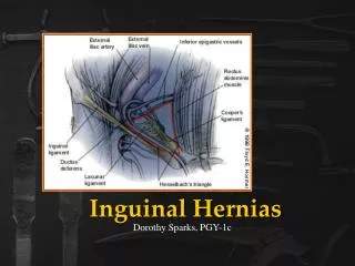
Anatomy of hernias. The tough abdominal wall confines the abdominal organs within the largest cavity in the body, extending from the diaphragm to the pelvic canal.

E N D
The tough abdominal wall confines the abdominal organs within the largest cavity in the body, extending from the diaphragm to the pelvic canal. • The arrangement of the aponeuroses of abdominal muscles, their attachments, and fibre direction are important to understand when attempting hernia reconstruction. Fibres of the tendinous aponeuroses of the external abdominal oblique, internal abdominal oblique, and transverse abdominal muscles converge and join on midline to form a thick white band, termed the linea alba, which is located between the rectus abdominis muscles. • The linea alba is widest in the cranial abdomen, and it narrows considerably before it attaches to the pubis by the prepubic tendon (sometimes termed the cranial pubic ligament). Fibers of the flat tendinous aponeuroses of abdominal wall muscles pass either superficially or deep to the rectus abdominis muscles that extend in a cranial and caudal direction from the first costal cartilage to the pecten of the pubis.
The external abdominal oblique aponeurosis always runs superficial to the rectus abdominis muscle. The location of the aponeurosis of the internal abdominal oblique varies along the length of the abdomen. In the cranial third of the abdominal wall, fibres pass both deep and superficial to the rectus abdominis muscle. From the umbilicus caudally, all of the internal abdominal oblique fibres run superficial to the rectus abdominis muscle. • Fibres of the transverse abdominal muscle pass deep to the rectus abdominis muscle in the cranial two thirds of the abdominal wall, but in the caudal third, fibres run superficial to the rectus abdominis muscle. • Whereas the external rectus sheath contains tendinous aponeuroses of the abdominal muscles that run superficial to the rectus abdominis muscle, the internal rectus sheath is composed of sheets of fascia that are deep to this muscle. The external rectus sheath has been shown to be the primary strength-holding layer throughout ventral midline closures
On the lateral aspect of the abdomen, fibers of the external abdominal oblique muscles, thoracodorsal fascia and extend in a caudoventral direction before giving rise to their broad aponeurosis. • Fibers of the internal abdominal oblique muscle, which lie immediately deep to the external abdominal oblique muscle, arise from the thoracolumbar fascia caudal to the last rib and from the tuber coxae. These fibers extend cranioventrally, crossing the more superficial fibers of the external abdominal oblique muscles at nearly right angles. • The deeper transverse abdominal muscle comes from two parts: the lumbar portion arises from the transverse processes of the lumbar vertebrae and the thoracolumbar fascia and the costal portion arises from the medial sides of the twelfth and thirteenth ribs and from the eighth to eleventh costal cartilages. Fibers from this muscle extend in a dorsoventral direction. The transverse fascia and peritoneum line the inner surface of this muscle.
Hernias • Hernia is a protrusion of the contents of a body cavity through a weak spot of the body wall. This may occur by accident or due to normal anatomical opening, which does not completely fulfil its physiological function. So, a part of an internal organ bulges through a weakened muscle, tissue, or membrane that would normally contain it. Hernias may be congenital or acquired. • Congenital hernias may involve the diaphragm or the abdominal wall. Hernias involving the diaphragm are of 3 main types: peritoneopericardial, in which abdominal contents are found extending into the pericardial sac; pleuroperitoneal, in which abdominal contents are found within the pleural cavity; and hiatal, in which the abdominal oesophagus, gastroesophageal junction, and/or portions of the stomach protrude through the oesophageal hiatus of the diaphragm into the thoracic cavity.
In acquired hernias, there is usually a history of trauma. • There is some genetic predisposition and some environmental factors that can lead to an increase of ruptures. Some of those environmental effects that could have an effect in the rate of hernias can be increased pressure at early age (piglet piling due to handling, cold temperature) or deficient castration technique. • Hernias can affect the growth of the animal, as well as its survival rate and the value of the carcass. • https://www.youtube.com/watch?v=M_iFDwP51VI
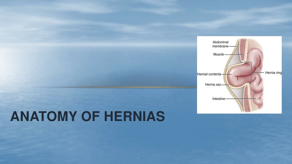
HERNIA ANATOMY • Hernia consist of 3 parts: • Hernial Ring: it's a rupture in muscles of the abdominal wall such as ventral hernia or widening of natural orifices such as the umbilicus, or may be a natural passageway such as the inguinal canal. • Hernial Sac: a fold of skin surrounding contents of hernia with muscle fibres and fibrous tissue in addition and peritoneum may be founded beneath the skin. In the cases of abdominal hernia, the sac consists of three parts the neck, that part closest to the ring and the bottom a lower part of the sac and the body that is between them. • Contents: the contents of the hernia differ depending on the site, may be part of the intestine called Enterocele or the presence of omentum is called Omentocele. In a few cases, content is part of the stomach is called Gastrocele or bladder Vesicocele.
Pathophysiology of hernias • The overall success of a hernia repair (and often the prognosis for the patient) rarely depends on the repair itself but more on management of the sequelae to organ herniation or internal damage from trauma that impair normal body function. • The severity of functional alteration depends on the cause, location, and content of the hernia. Important, often life-threatening, sequelae associated with abdominal hernias can be attributed to space-occupying effects (known as loss of domain), incarceration or obstruction, or strangulation. • The clinical condition of the animal at presentation, whether the hernia is open to the outside or not, concurrent injuries to distant structures, and organ compromise from the trauma must also be factored when attempting to predict patient outcome