Download
1 / 19
190 likes | 311 Views
Effects of Animal Viruses on Host Cells. (What does the virus do to the host cell?). Morphological Changes. Virus lytic infections often cause very distinctive, visible changes in the infected cell. These changes are called cytopathic effects (CPE) and they include:

E N D
Effects of Animal Viruses on Host Cells (What does the virus do to the host cell?)
Morphological Changes • Virus lytic infections often cause very distinctive, visible changes in the infected cell. These changes are called cytopathic effects (CPE) and they include: • Inclusion bodies – microscopically these are visible sites of viral assembly or cellular damage. They are often used as a diagnostic tool. Examples include: • Virions in the nucleus (Adenovirus) • Virions in the cytoplasm (Rhabdovirus- negri bodies of rabies virus)
Morphological changes • Viral protein associated with host microtubules (Reovirus) • Factories of viral replication in the cytoplasm (Poxvirus) • Clumps of ribosomes in capsids (Arenavirus) • Clumps of chromatin (herpesviruses) • Morphological alterations • Nuclear pyknosis (shrinking) (Picornaviruses) • Proliferation of membranes (Picornaviruses) • Proliferation of the nuclear membrane (Alphaviruses) • Formation of vacuoles in the cytoplasm (Papovaviruses) • Apoptosis (will discuss this more later) • Formation of syncytia (Paramyxoviruses and Coronaviruses) which are giant, multinucleated cells formed by the fusion of plasma membranes
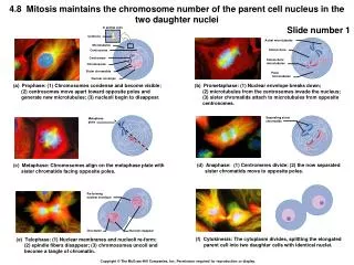
Morphological changes • Margination and breakage of chromosomes (Herpesviruses) • Rounding up and detachment of tissue culture cells – due to apoptosis (Herpes and Rhabdoviruses)
Morphological changes • CPE is very rarely caused by a harmful protein with no other purpose in the infective process. • CPE is usually a secondary result of changes in the host metabolism caused by viral replication. • Viruses may halt or alter host cell DNA synthesis, transcription, and/or protein synthesis (translation)
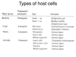
Changes in the Host Cell Due To Viral Infection • Eukaryotic viruses are more likely to target host cell translation rather than transcription or DNA replication because the half-life of mRNAs is so long in eukaryotic cells (T1/2=600 minutes). • If the virus wants to use the host translation machinery, it will need to compete with host mRNAs for a long time. • Therefore, it needs to shut off or slow down host mRNA translation while allowing viral translation to continue. • There are three steps in protein synthesis (translation) that are potential targets for viral action.
Inhibition of Eukaryotic Protein Synthesis • Initiation – requires mRNA, 2 ribosomal subunits, special met-tRNAmet, initiation factors (eIF 1-6), ATP, and GTP • Elongation – requires charged tRNAs, elongation factors (EF1, and EF1B), and GTP • Termination – requires a termination factor and GTP • Mechanisms to inhibit host cell protein synthesis may vary for the same virus in different hosts. They include: • Competition • The virus may produce an overabundance of mRNAs an/or produce viral products that actually bind to and tie up host cell mRNAS to make them inaccessible to ribosomes (rhabdovirus and reovirus).
Inhibition of Eukaryotic Protein Synthesis • The viral mRNAs may have highly accessible and easily recognized initiation sequences that allow them to compete more effectively for the initiation factor 2 (eIF2). Therefore, there is a higher initiation of translation of the viral proteins (mengovirus and influenza virus). • There may be an inhibition of transport of host mRNA from the nucleus. Adenovirus allows selective transport of viral mRNAs from the nucleus, while inhibiting the transport of the host mRNAs. • The virus may degrade the host mRNAs • Bunyavirus uses primers derived from host cell mRNA for its own transcription in the cytoplasm. The 5’ cap of the host mRNA plus 10-14 bases are cleaved by a viral enzyme. This is then used as the primer for transcription. This is similar to what influenza virus does in thenucleus.
Inhibition of Eukaryotic Protein Synthesis • Herpes viruses (remember that they control the timing of their gene expression through positive and negative feedback loops) • First host mRNA is degraded. This is mediated by a protein found as part of the virion • Next, at the time that the genes get expressed, there is a complete shut down of host protein synthesis. This is due to a newly synthesized viral protein. • Blockage of initiation complex formation • Pox virus alters the specificity of the host cell ribosomes so that the viral mRNA is preferentially recognized • A phosphoprotein that is part of the virion acts on the 40S ribosomal subunit to hinder its association with the met-tRNA met, GTP, and eIF2. This equally inhibits host and viral translation.
Inhibition of Eukaryotic Protein Synthesis • Another viral protein that is not a part of the virion, somehow allows specific translation of viral messages only. • Covalent modification of translation related components • Inactivation by cleavage – Poliovirus cleaves the cap-binding complex (eIF4) so that capped host mRNAs are no longer translated. How is viral mRNA then translated? • Inactivation by phosphorylation • Influenza A virus and adenoviruses inactivate the cap binding reaction by removing the required phosphate from eIF-4E • Picornaviruses act indirectly on the cap binding complex by phosphorylating 4E-bp1 which then competes with eIF-4G for binding to the active form of eIF-4E
Inhibition of Eukaryotic Protein Synthesis • A poxvirus structural core protein sequentially phosphorylates and inactivates three ribosomal proteins • VSV, adenovirus, mengovirus, and reovirus phosphorylate the subunit of eIF2. When eIF2 is phosphorylated, it forms a complex with GEF (GTP exchanging factor or eIF-2B) that can’t exchange GDP for GTP. The exchange of GDP for GTP normally occurs upon formation of the initiation complex. This exchange allows to participate in the formation of a new initiation complex. When the exchange does not occur, is blocked from participating in the formation of a new initiation complex and the rate of translation initiation is slowed down for both host and viral mRNA. Since there is an overabundance of viral mRNA, this tips the scale even more in favor of viral mRNA translation.
Inhibition of Eukaryotic Protein Synthesis • The enzyme that phosphorylates eIE2 is called protein kinase RNA dependent (PKR). Its mechanism of action will be discussed later. • Increases in intracellular cation concentrations – one of the environmental conditions that can influence the rate or efficiency of an enzymatic reaction is the concentration of particular cations (Na+, K+). • Some viruses alter the membrane permeability to cause an increase in intracellular Na+. • This inhibits host cell, but not viral translation. (Sindbis virus inhibits the Na+ pump in the plasma membrane).
Inhibition of Eukaryotic Protein Synthesis • Some viruses produce inhibitory proteins • Mengoviruses produce a protein that binds to ribosomes and blocks translation after formation of the initiation complex, i.e., it inhibits elongation. • In summary, as an example, mengoviruses may use three different methods to halt host cell protein synthesis: • Competition for initiation complexes • Inhibition of elongation • Phosphorylation of eIF-2. • The viral RNA of this virus is so efficient in initiation that it must use the other two mechanisms to free viral RNA for packaging into the genome!
Changes in the Host Cell Due To Viral Infection • Inhibition of host cell transcription • This may be secondary to the shut off of host mRNA translation. • Factors required for host cell transcription are not replaced when translation is halted. • Some viruses appear to actually inhibit host cell mRNA synthesis by acting directly or indirectly on RNA polymerase II by unknown mechanisms (polio and reovirus) • The viral RNA may inhibit transcription – there is evidence that the VSV leader sequence may bind to an initiation factor for host transcription making it unavailable for host transcription.
Changes in the Host Cell Due To Viral Infection • Inhibition of host cell DNA replication • This may be secondary to decreased translation • Some viruses have proteins that may specifically inhibit host DNA synthesis (reovirus) • Poxvirus produces a protein that degrades SS DNA at the replication fork • Herpesviruses displace host chromatin from its normal association with nuclear matrix proteins