Download
1 / 28
320 likes | 460 Views
CS/BIO 471/671 – Algorithms for Bioinformatics. The Structure and Functions of Proteins. The many functions of proteins. Mechanoenzymes: myosin, actin Rhodopsin: allows vision Globins: transport oxygen Antibodies: immune system Enzymes: pepsin, renin, carboxypeptidase A

E N D
CS/BIO 471/671 – Algorithms for Bioinformatics The Structure andFunctions of Proteins
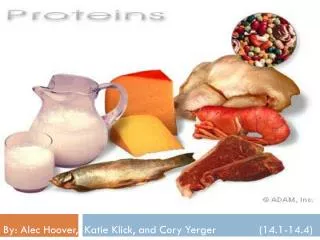
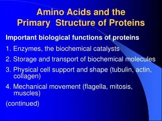
The many functions of proteins • Mechanoenzymes: myosin, actin • Rhodopsin: allows vision • Globins: transport oxygen • Antibodies: immune system • Enzymes: pepsin, renin, carboxypeptidase A • Receptors: transmit messages through membranes • Vitelogenin: molecular velcro • And hundreds of thousands more… Protein Structure and Function
Complex Chemistry Tutorial • Molecules are made of atoms! • There is a lot of hydrogen out there! • Atoms make a “preferred” number of covalent (strong) bonds • C – 4 • N – 3 • O, S – 2 • Atoms will generally “pick up” enough hydrogens to “fill their valence capacity”in vivo. • Molecules also “prefer” to have a neutral charge Protein Structure and Function
Biochemistry • In the context of a protein… • Oxygen tends to exhibit a slight negative charge • Nitrogen tends to exhibit a slight positive charge • Carbon tends to remain neutral/uncharged • Atoms can “share” a hydrogen atom, each making “part” of a covalent bond with the hydrogen • Oxygen: H-Bond donor or acceptor • Nitrogen: H-Bond donor • Carbon: Neither Protein Structure and Function
Proteins are chains of amino acids • Polymer – a molecule composed of repeating units Protein Structure and Function
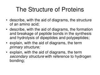
R O H N C C OH H H Amino acid composition • Basic Amino AcidStructure: • The side chain, R,varies for each ofthe 20 amino acids Side chain Aminogroup Carboxylgroup Protein Structure and Function
The Peptide Bond • Dehydration synthesis • Repeating backbone: N–C –C –N–C –C • Convention – start at amino terminus and proceed to carboxy terminus O O Protein Structure and Function
Peptidyl polymers • A few amino acids in a chain are called a polypeptide. A protein is usually composed of 50 to 400+ amino acids. • Since part of the amino acid is lost during dehydration synthesis, we call the units of a protein amino acid residues. amidenitrogen carbonylcarbon Protein Structure and Function
Side chain properties • Recall that the electronegativity of carbon is at about the middle of the scale for light elements • Carbon does not make hydrogen bonds with water easily – hydrophobic • O and N are generally more likely than C to h-bond to water – hydrophilic • We group the amino acids into three general groups: • Hydrophobic • Charged (positive/basic & negative/acidic) • Polar Protein Structure and Function
The Hydrophobic Amino Acids Proline severely limits allowable conformations! Protein Structure and Function
The Charged Amino Acids Protein Structure and Function
The Polar Amino Acids Protein Structure and Function
More Polar Amino Acids And then there’s… Protein Structure and Function
Planarity of the peptide bond Psi () – the angle of rotation about the C-C bond. Phi () – the angle of rotation about the N-C bond. The planar bond angles and bond lengths are fixed. Protein Structure and Function
Phi and psi • = = 180° is extended conformation • : C to N–H • : C=O to C C=O C N–H Protein Structure and Function
The Ramachandran Plot • G. N. Ramachandran – first calculations of sterically allowed regions of phi and psi • Note the structural importance of glycine Observed (non-glycine) Observed (glycine) Calculated Protein Structure and Function
Primary & Secondary Structure • Primary structure = the linear sequence of amino acids comprising a protein:AGVGTVPMTAYGNDIQYYGQVT… • Secondary structure • Regular patterns of hydrogen bonding in proteins result in two patterns that emerge in nearly every protein structure known: the -helix and the-sheet • The location of direction of these periodic, repeating structures is known as the secondary structure of the protein Protein Structure and Function
The alpha helix 60° Protein Structure and Function
Properties of the alpha helix • 60° • Hydrogen bondsbetween C=O ofresidue n, andNH of residuen+4 • 3.6 residues/turn • 1.5 Å/residue rise • 100°/residue turn Protein Structure and Function
Properties of -helices • 4 – 40+ residues in length • Often amphipathic or “dual-natured” • Half hydrophobic and half hydrophilic • Mostly when surface-exposed • If we examine many -helices,we find trends… • Helix formers: Ala, Glu, Leu,Met • Helix breakers: Pro, Gly, Tyr,Ser Protein Structure and Function
The beta strand (& sheet) 135° +135° Protein Structure and Function
Properties of beta sheets • Formed of stretches of 5-10 residues in extended conformation • Pleated – each C a bitabove or below the previous • Parallel/aniparallel,contiguous/non-contiguous Protein Structure and Function
Parallel and anti-parallel -sheets • Anti-parallel is slightly energetically favored Anti-parallel Parallel Protein Structure and Function
Turns and Loops • Secondary structure elements are connected by regions of turns and loops • Turns – short regionsof non-, non-conformation • Loops – larger stretches with no secondary structure. Often disordered. • “Random coil” • Sequences vary much more than secondary structure regions Protein Structure and Function
Levels of Protein Structure • Secondary structure elements combine to form tertiary structure • Quaternary structure occurs in multienzyme complexes • Many proteins are active only as homodimers, homotetramers, etc.
Protein Structure Examples Protein Structure and Function
Views of a protein Wireframe Ball and stick Protein Structure and Function
Views of a protein Spacefill Cartoon CPK colors Carbon = green, black, or grey Nitrogen = blue Oxygen = red Sulfur = yellow Hydrogen = white Protein Structure and Function