Download
1 / 29
290 likes | 407 Views
The Auditory Periphery. I . Introduction to hearing - Sound is a pressure wave, and pure tones can be characterized by amplitude (measured indirectly as dB) and frequency - Dynamic range of hearing in human is about 0-100 dB (whisper to pain) and 20-20K Hz. - Types of damage to ear

E N D
I. Introduction to hearing - Sound is a pressure wave, and pure tones can be characterized by amplitude (measured indirectly as dB) and frequency - Dynamic range of hearing in human is about 0-100 dB (whisper to pain) and 20-20K Hz.
- Types of damage to ear - Chronic severely loud sound exposure kills hair cells - Ototoxic antibiotics, e.g. streptomycin or gentamicin, kills hair cells - Hearing loss of high frequencies with age, aka presbycusis.
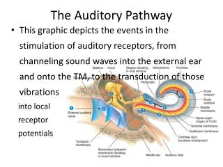
I. Anatomy of the ear A) Outer ear - Pinna: collects and funnels sound toward the opening to the auditory canal, thus amplifying sound; sound shadows from sounds coming from different positions in space provide resolution of sound origin - External auditory canal: acts as resonant tube for 4 kHz tones.
B) Middle ear - Impedance matching of air to water sound transmission by three mechanisms (1) Area of tympanic membrane is 20x that of stapes footplate (2) Lever advantage of ossicles (malleus, incus, stapes) (3) Oval window of stapes is smaller than eardrum.
- Middle ear muscles (tensor tympani, stapedius): restrict ossicle movement in response to loud sound to prevent damage. The tensor tympani reduces the area of the flexible membrane in the eardrum; the stapedius restricts the amplitude of stapes motion.
- Damage in the middle ear is called “conductive.” A common cause is otosclerosis in which bony growth impedes the movement of the middle ear ossicles.
C) Inner ear (more on cochlea below) - Damage in the inner ear is called “sensorineural” - One can distinguish between conductive and sensorineural hearing loss by comparing the audibility of a tuning fork held in the air, or pressed against the skull.
In conductive hearing loss, the latter position is effective at presenting sound by bone conduction, thus overcoming the conductive loss that pertains to air-borne sound.
III. The Cochlea - The stapes footplate moving in and out if the oval window sets up a fluid wave within the scala vestibule. The motion of this wave causes the cochlear partition to move alternatively up and down within the cochlear duct.
The cochlear partition of the basilar membrane and the hair cells, supporting cells and extracellular structures lying upon it, and the overlying tectorialmembrane. These all lie just under the scala media, the central membraneous tube flanked on top and bottom by the scala vestibule and scala tympani, respectively.
The scala media is bounded by Reisnner’s membrane on the top and is actually delimited by the apical surface of the hair cell epithelium on the bottom, not by the basilar membrane. This distinction is important because the scala media is filled with endolymph (high K, low Na), which is produced by a secreting epithelium called the striavascularis that lines the outer wall of scala media.
The scalavestibule and tympani contain perilymph (low K, high Na). Since the basilar membrane itself does not present a diffusion barrier, the basal surface of the hair cells is bathed by low K perilymph while their apical, hair-bearing surface faces high K endolymph. This disposition of ionic media is important for hair cell function.
Since endolymph is 80 mV positive to perilymph, this endolymphic potential provides additional driving force for K+ ions to flow into hair cells. - Meniere’s disease arises under conditions in which the normal circulation of endolymp is blocked or altered resulting in sensorineural hearing loss.
- There are two classes, inner and outer, of hair cells. In a cochlear cross section one can see the single row of flask-shaped inner hair cells, and three rows of columnar outer hair cells.
- Stereocilia of hair cells are arranged in rows of increasing height and are embedded in the tectorial membrane; shear of basilar membrane with respect to tectorial membrane causes displacement of hair bundle.
- Tip links join nonselective cation channels on one stereocilium to the next tallest one tilting of the stereocilia causes channel to open or close open channel allows K, the primary cation of endolymph to travel down steep electrical gradient into hair cell and depolarize it if depolarized, hair cell opens voltage gated Ca channels, allows Ca in, and releases Glu onto the nerve ending.
- Hair cell adaptation may occur through two suggested negative feedback mechanisms (1) end of tip link is attached to myosin-like motor (myosin 1C) on stereocilia, movement up or down changes the set point of system. Myosin 1C exerts steady force on the transduction channel.
(2) Ca entry feeds back to regulate the mechanotransduction channel - Synaptic transmission: At its basal end the hair cell is contacted by afferent fibers whose cell bodies are located in the spiral ganglion and which send an axon centrally to end on the cochlear nucleus of the brainstem.
When the hair cell is depolarized by opening of the transducer channel, voltage dependant calcium channels in the basolateral membrane are opened and synaptic vesicles fuse with plasma membrane to release Glu, which binds to AMPA receptors causing excitation of the afferent axon.
This depolarizing effect of v-gated Ca+ influx is counterbalanced by v-gated K+ channels that dominate the basolateral conductance of the hair cell and allow outward flow of K into low K perilymph. - Individual afferent neurons makes only a single contact with a single inner hair cell.
At the point of contact, the hair cell places a synaptic ribbon, an electron-dense body to which a number of transmitter-containing vesicles are tethered. It is thought the ribbon serves to motivate vesicles for release during stimulation by sound and during the ongoing spontaneous activity that occurs in silence.
- Nearly all afferent nerves end on inner hair cells, whereas efferents end on outer hair cells. The crossed olivo-cochlear bundle arises from the contralateral superior olivary complex and synapses massively on outerhair cells. This pathway is inhibitory and when activated, causes a loss of sensitivity and frequent selectivity in afferent fiber response.
- Outer hair cells exhibit electromotility (contract on depolarization and vice versa) to regulate distance of tectorial membrane to inner hair cells and adjust their sensitivity; this mechanism depends on a motor protein called prestin.
Targeted disruption of prestin in mice causes deafness. - OHCs express a Ca channel opened by ACh from efferent nerves; the Ca causes K channels to open and results in hyperpolarization, which ultimately results in loss of sensitivity.
- Otoacoustic emissions: active movement of basilar membrane can generate sound. - Active motions of OHCs are driven by the sound-evoked change in membrane potential. These active motions enhance the overall vibration pattern of the cochlear partition, thereby causing larger deflections of the IHC bundle.
Cochlear signaling is enhanced by OHC electromotility. If OHCs are damaged, the cochle becomes much less sensitve.
Thus, the efferent innervation of the cochlea, which causes a suppression of cochlear sensitivity, must do so by inhibiting OHCs. This occurs via cholinergic inhibition of cochlear hair cells from olivocochlear efferent activity.
Hair cells are depolarized by nicotinic AChR, but only briefly. Ca++ enters the cell thru the receptor and causes the opening of Ca++ dependant K+ channels, of which there are many, causing a net hyperpolarization of the hair cell’s membrane.