Download
1 / 37
430 likes | 1.25k Views
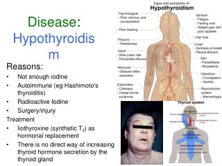
Thyroid Eye Disease & Other Orbital Conditions. March 11, 2008. Thyroid Eye Disease: . Thyrotoxicosis (Graves’ Disease) Autoimmune disorder Third or Fourth decade Women > Men Euthyroid Graves disease – Eye signs without clinical hyperthyroid (5%). Pathogenesis of Thyroid Eye Disease:.

E N D
Thyroid Eye Disease&Other Orbital Conditions March 11, 2008
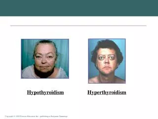
Thyroid Eye Disease: • Thyrotoxicosis (Graves’ Disease) • Autoimmune disorder • Third or Fourth decade • Women > Men • Euthyroid Graves disease – Eye signs without clinical hyperthyroid (5%).
Pathogenesis of Thyroid Eye Disease: • IgG infiltrates the EOM resulting in cellular infiltration into the muscle belly. • Muscle fiber fibrosis leading to restrictive myopathy & diplopia. • Inflammatory cellular infiltration • Increase in the orbital contents
Clinical Manifestations of Thyroid Eye Disease: 1) Soft Tissue Involvement: • Periorbital & Lid edema • Conjunctival & episcleral hyperemia • may outline the horizontal rectus muscles. • Chemosis • Dry eye (infiltration of the lacrimal gland).
Clinical Manifestations of Thyroid Eye Disease: 2) Proptosis • An abnormal protrusion of the globe. • unilateral or bilateral • symmetrical or asymmetrical • frequently permanent
Measuring Proptosis • Measured with an exophthalmometer • Hertel • Readings greater than 20 mm are indicative of proptosis. • A difference of 2 mm is suspicious. • Normal values depends on race & gender.
Pseudo-proptosis: • Facial asymmetry • High myopia or buphthalmos • Lid retraction • Enophthalmos
Clinical Manifestations of Thyroid Eye Disease: 3) Lid Retraction: • Fibrosis of the levator. • Sympathetic overstimulation of Mueller’s muscle. • Dalrymple sign- lid retraction in primary gaze. • Von Graefe sign- retraction on downgaze • Kocher sign- stare with attentive fixation
Clinical Manifestations of Thyroid Eye Disease: 4) Optic Neuropathy: • Affecting about 5% of patients with thyroid eye disease • Compression of the optic nerve by the enlarged EOM’s at the orbital apex. • What signs may be involved?
Clinical Manifestations of Thyroid Eye Disease: 5) Restrictive myopathy • Affects 30 to 50% of patients with thyroid eye disease. • IOP increase on upgaze (> 4 mmHg) due to fibrotic inferior rectus. • Resistance to retrodisplacement (multiple fibrotic EOM’s) • Absence of Bell’s phenomenon (inferior rectus) • Occurs first due to edema and later due to fibrosis.
Clinical Work-up of the Suspected Graves Disease Patient • VA • Exophthalmometry • Resistance to Retrodisplacement of Globe • An Absent Bell’s Phenomenon • Lagophthalmos • Slit lamp • Schirmer’s • Visual Field Testing
Imaging Work-up of the Suspected Graves Disease Patient • Looking for the enlarged EOM’s • Can use ultrasound, CT or MRI. • MRI better for soft tissue imaging.
Classifying Graves Disease (Remember NO SPECS) Class 0 No physical signs or symptoms 1 Only signs 2 Soft tissue involvement 3 Proptosis > 3mm 4 EOM involvement 5 Corneal involvement 6 Sight loss
Carotid-cavernous fistula: • Communication between the carotid artery and the cavernous sinus. • Venous and arterial stasis around the eye and orbit.
Mucocele: • A slowly expanding cystic accumulation of mucoid secretions. • Normal sinus drainage is obstructed • Follows infections, allergies, sinusitis
Dacryocystitis vs. Mucocele • Mucocele – A cystic mass usually filled with mucous. Typically arises after sinusitis.
Encephalocele: • Herniation of intracranial contents through a congenital defect of the base of the skull. • Usually seen in infancy • Typically involves the superomedial part of the orbit.
Cavernous hemangioma: • Most common benign orbital tumor in adults. • Most common in women • Slowly progressive & unilateral • Growth is accelerated during pregnancy • Tumor usually located within the muscle cone.
Preseptal vs. OrbitalCellulitis: Both Conditions show: • Chemosis • Conjunctival injection • Pain • Redness • Swelling of the lids
Preseptal: No Proptosis F.R.O.M. of EOM’s Normal Pupils Normal Visual Acuity Orbital: Proptosis Ophthalmoplegia + APD Reduced Visual Acuity Preseptal vs. Orbital Cellulitis: