Download
1 / 26
260 likes | 348 Views
Making an Intraop Neuropathology preparation. Starring Ed Plowey. Surgeon obtains specimen. Tissue within needle is gently teased onto saline MOISTENED (not soaked) Telfa. Set up. Instruments: Clean surface, Scalpel, Fine forceps, labeled slides, OPEN Fixative jar. Specimen arrival.

E N D
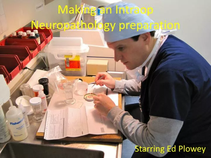
Making an Intraop Neuropathology preparation Starring Ed Plowey
Surgeon obtains specimen • Tissue within needle is gently teased onto saline MOISTENED (not soaked) Telfa
Set up • Instruments: Clean surface, Scalpel, Fine forceps, labeled slides, OPEN Fixative jar
Specimen arrival • Labeling slides
Touch Prep • Either: • Turn slide frosted side down and touch to entire specimen Or if specimen firm (e.g. meningioma) • Pick up with forceps and dab twice to slide • DO NOT SLIDE OR SWIRL • FIX IMMEDIATELY • i.e. ASAP • i.e. in less than 1 second
Touch Prep Turn slide frosted side down and touch entire specimen • FIX IMMEDIATELY i.e. ASAP i.e. < 1 second
Touch Prep Video • http://www.youtube.com/watch?v=Koxv1L5gyuQ
Pituitary adenoma TP Slide overview of Adenoma TP 20X of adenoma Pituitary adenomas are diagnosed intra-operatively by Touch Prep ONLY
Smearing specimen size • No Larger than tip of pencil • 0.5mm3
Smearing • Slides can be held parallel or perpendicular (as shown here)
Smearing Video • http://www.youtube.com/watch?v=7BFWS6Izdhk
Examples of good smears Useable area 2 parts of specimen on one slide Superb Smear
Summary: Set up: Key features • Slides labeled • Specimen on moist Telfa pad • Not Dry: Not immersed in saline • IF specimen uniform • Cut off piece up to (but not exceeding) size of tip of pencil (<0.5mm3) • IF specimen is biphasic • Make two slides or • Place small piece on opposite sides of slide
Common problems / solutions • Too much tissue on slide • Use less • Air drying • Be sure fixative jar (95% ethanol) is open and adjacent to prep site • Drop slide into jar for fastest fixation • No tissue touches off or smears • Move on to frozen section
Smear problems: Too much tissue • Solution: Obviously use less
TP/Smear Problems: Air dried • Solution: Have fixative jar open and closer before starting prep
Not enough material all at bottom of slide • Solution: Start by placing specimen ½” from frosted end of slide
Specimen not touching off • Try smear and if that doesn’t work go to frozen
What is wrong with this picture? No Gloves !