Download
1 / 1
10 likes | 101 Views
Sleep Apnea Induced Sleep Cycle Fragmentation David Whisler Knapp Center For Biomedical Discovery University of Chicago Fahed Hakim, M.D. The Effect of TRIF and MYD88 Knockout Genes on Tumor Size in Sleep Fragmented Mice.

E N D
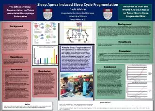
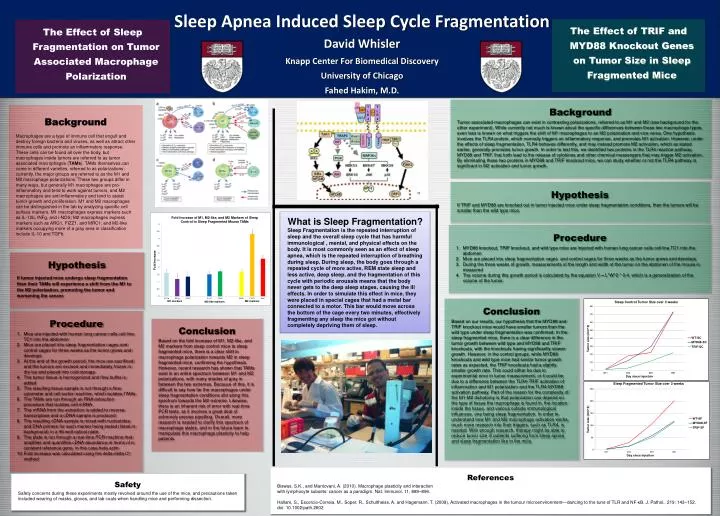
Sleep Apnea Induced Sleep Cycle Fragmentation David Whisler Knapp Center For Biomedical Discovery University of Chicago Fahed Hakim, M.D. The Effect of TRIF and MYD88 Knockout Genes on Tumor Size in Sleep Fragmented Mice The Effect of Sleep Fragmentation on Tumor Associated Macrophage Polarization Background Tumor associated macrophages can exist in contrasting polarizations, referred to as M1 and M2 (see background for the other experiment). While currently not much is known about the specific differences between these two macrophage types, even less is known on what triggers the shift of M1 macrophages to an M2 polarization and vice versa. One hypothesis involves the TLR4 protein, which normally triggers an inflammatory response, and promotes M1 activation. However, under the effects of sleep fragmentation, TLR4 behaves differently, and may instead promote M2 activation, which as stated earlier, generally promotes tumor growth. In order to test this, we identified two proteins in the TLR4 reaction pathway, MYD88 and TRIF, that both lead to the release of cytokines and other chemical messengers that may trigger M2 activation. By eliminating these two proteins in MYD88 and TRIF knockout mice, we can study whether or not the TLR4 pathway is significant in M2 activation and tumor growth. Background Macrophages are a type of immune cell that engulf and destroy foreign bacteria and viruses, as well as attract other immune cells and promote an inflammatory response. These cells can be found all over the body, but macrophages inside tumors are referred to as tumor associated macrophages (TAMs). TAMs themselves can come in different varieties, referred to as polarizations: currently, the major groups are referred to as the M1 and M2 macrophage polarizations. These two groups differ in many ways, but generally M1 macrophages are pro-inflammatory and tend to work against tumors, and M2 macrophages are anti-inflammatory and tend to assist tumor growth and proliferation. M1 and M2 macrophages can be distinguished in the lab by analyzing specific cell surface markers. M1 macrophages express markers such as IL-12b, INFg, and i-NOS; M2 macrophages express markers such as ARG1, FIZZ1, and MRC1; and M2-like markers occupying more of a gray area in classification include IL-10 and TGFb. Hypothesis If TRIF and MYD88 are knocked out in tumor injected mice under sleep fragmentation conditions, then the tumors will be smaller than the wild type mice. What is Sleep Fragmentation? Sleep Fragmentation is the repeated interruption of sleep and the overall sleep cycle that has harmful immunological , mental, and physical effects on the body. It is most commonly seen as an effect of sleep apnea, which is the repeated interruption of breathing during sleep. During sleep, the body goes through a repeated cycle of more active, REM state sleep and less active, deep sleep, and the fragmentation of this cycle with periodic arousals means that the body never gets to the deep sleep stages, causing the ill effects. In order to simulate this effect in mice, they were placed in special cages that had a metal bar connected to a motor. This bar would move across the bottom of the cage every two minutes, effectively fragmenting any sleep the mice got without completely depriving them of sleep. Procedure MYD88 knockout, TRIF knockout, and wild type mice are injected with human lung cancer cells cell line TC1 into the abdomen Mice are placed into sleep fragmentation cages and control cages for three weeks as the tumor grows and develops During the three weeks of growth, measurements of the length and width of the tumor on the abdomen of the mouse is measured The volume during this growth period is calculated by the equation V = L*W^2 * 0.4, which is a generalization of the volume of the tumor. Hypothesis If tumor injected mice undergo sleep fragmentation then their TAMs will experience a shift from the M1 to the M2 polarization, promoting the tumor and worsening the cancer. Conclusion Based on our results, our hypothesis that the MYD88 and TRIF knockout mice would have smaller tumors than the wild type under sleep fragmentation was confirmed. In the sleep fragmented mice, there is a clear difference in the tumor growth between wild type and MYD88 and TRIF knockouts, with the knockouts having significantly slower growth. However, in the control groups, while MYD88 knockouts and wild type mice had similar tumor growth rates as expected, the TRIF knockouts had a slightly smaller growth rate. This could either be due to experimental error in tumor measurement, or it could be due to a difference between the TLR4-TRIF activation of inflammation and M1 polarization and the TLR4-MYD88 activation pathway. Part of the reason for the complexity of the M1-M2 dichotomy is that polarization can depend on the type of tissue the macrophage is found in, the location inside the tissue, and various outside immunological influences, one being sleep fragmentation. In order to understand how M1 and M2 macrophage activation works, much more research into their triggers, such as TLR4, is needed. With enough research, therapy might be able to reduce tumor size in patients suffering from sleep apnea and sleep fragmentation like in the mice. Procedure Mice are injected with human lung cancer cells cell line TC1 into the abdomen Mice are placed into sleep fragmentation cages and control cages for three weeks as the tumor grows and develops At the end of the growth period, the mice are sacrificed and the tumors are excised and immediately frozen in dry ice and placed into cold storage The tumor tissue is homogenized and flow buffer is added The resulting tissue sample is run through a flow cytometer and cell sorter machine, which isolates TAMs The TAMs are run through an RNA extraction procedure that isolates cell mRNA The mRNA from the extraction is added to reverse transcriptase and a cDNA sample is produced The resulting cDNA sample is mixed with nucleotides and DNA primers for each marker being tested (listed in background) in a 96 well optical plate The plate is run through a real-time PCR machine that amplifies and quantifies cDNA abundance in terms of a constant reference gene, in this case beta actin Fold increase was calculated using the delta-delta Ct method Conclusion Based on the fold increase of M1, M2-like, and M2 markers from sleep control mice to sleep fragmented mice, there is a clear shift in macrophage polarization towards M2 in sleep fragmented mice, confirming the hypothesis. However, recent research has shown that TAMs exist in an entire spectrum between M1 and M2 polarizations, with many shades of gray in between the two extremes. Because of this, it is difficult to say how far the macrophages under sleep fragmentation conditions slid along this spectrum towards the M2 extreme. Likewise, there is an inherent risk of error with real-time PCR tests, as it involves a great deal of extremely precise pipetting. Overall, more research is needed to clarify this spectrum of macrophage states, and in the future learn to manipulate this macrophage plasticity to help patients. References Biswas, S.K., and Mantovani, A. (2010). Macrophage plasticity and interaction with lymphocyte subsets: cancer as a paradigm. Nat. Immunol. 11, 889–896. Hallam, S., Escorcio-Correia, M., Soper, R., Schultheiss, A. and Hagemann, T. (2009), Activated macrophages in the tumour microenvironment—dancing to the tune of TLR and NF-κB. J. Pathol., 219: 143–152. doi: 10.1002/path.2602 Safety Safety concerns during these experiments mostly revolved around the use of the mice, and precautions taken included wearing of masks, gloves, and lab coats when handling mice and performing dissection.