Download
1 / 43
550 likes | 950 Views
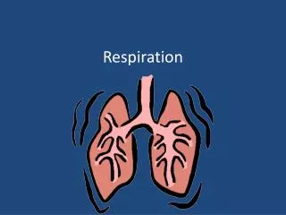
Respiration. Chapter 32 Pages. Why Exchange Gases?. The act of breathing is called respiration Cellular respiration converts the energy in nutrients into the ATP used by cells, requires oxygen and generates carbon dioxide as waste

E N D
Respiration Chapter 32 Pages
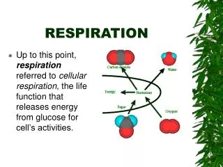
Why Exchange Gases? • The act of breathing is called respiration • Cellular respiration converts the energy in nutrients into the ATP used by cells, requires oxygen and generates carbon dioxide as waste • The circulatory system works in with the respiratory system • The circulatory system extracts oxygen from the air in your lungs, carries it within diffusing distance of each cell, then picks up carbon dioxide for release from the lungs • Cellular respiration depletes O2 levels, creating a concentration gradient that favors the diffusion of CO2 out of cells and the diffusion of O2 into them
Requirements for Diffusion • Animal respiratory systems are diverse but all meet three requirements that facilitate diffusion • Respiratory surfaces remain moist so gases can diffuse across cell membranes • Cells lining respiratory surfaces are very thin, to facilitate diffusion of gases through them • Respiratory systems have a sufficiently large area in contact with the environment to allow adequate gas exchange
Evolutionary Adaptations for Gas Exchange • Some animals in moist environments lack specialized respiratory structures • The outside of the body is covered by a thin, gas-permeable skin, which provides an adequate surface area for the diffusion of gases • If the body is small and elongated (microscopic roundworms) gases need to diffuse only a short distance to reach all cells • An animal’s body may be thin and flattened, (flatworms) most cells are close to the moist skin
Invertebrate Gas Exchange • The slow rate of gas exchange by diffusion may suffice for a larger, thicker bodied organism if energy demands are low, as sea jellies, which can be large but require little O2 • Another adaptation for gas exchange involves bringing the watery environment close to each cell - Sponges circulate seawater through channels within their bodies
Earthworms • For O2 delivery to cells, some animals combine a large skin surface area with well-developed circulation • In earthworms, gases diffuse through moist skin and are distributed throughout the body by a circulatory system • Blood in the skin capillaries rapidly carries off O2 that has diffused through the skin, maintaining a concentration gradient that favors the inward diffusion of oxygen • The worm’s elongated shape ensures a surface area relative to its internal volume
Respiratory Systems • Facilitate gas exchange by diffusion • Most animals have evolved specialized respiratory systems that interface with circulatory systems to exchange gases between their cells and the environment • Transfer of gases between environment and body cells usually occurs in stages that alternate between bulk flow and diffusion • During bulk flow, liquids or gases move through large spaces, from areas of higher to lower pressure • This contrasts with diffusion, where molecules move individually from higher to lower concentrations
Gas Exchange Occurs in Stages • For animals with well-developed respiratory systems • Air or water moves past a respiratory surface by bulk flow (down a pressure gradient), this is usually facilitated by muscular movements such as breathing • O2 and CO2 are exchanged through the respiratory surface by diffusion • O2diffuse into the capillaries of the circulatory system • CO2diffuses out • Gases are transported between the respiratory system and tissues by the bulk flow of blood as it is pumped throughout the body • Gases are exchanged between tissues and the circulatory system by diffusion; at the tissue level, O2 moves out of the capillaries into tissues, and CO2 moves from the tissues to the capillaries
Gas Exchange in Mammals Oxygenated blood Gases move in and out of the lungs by breathing 1 Deoxygenated blood CO2 O2 2 O2 and CO2 are exchanged in the lungs by diffusion O2 O2 CO2 O2 O2 alveoli (air sacs) Gases dissolved in the blood are transported by the circulatory system 3 left atrium right atrium right ventricle left ventricle O2 CO2 CO2 O2 and CO2 are exchanged in the tissues by diffusion 4 CO2 CO2 CO2
Gas Exchange in Aquatic Environments • Gills are the respiratory structures of some aquatic animals • The simplest gills (amphibian) are thin projections of the body surface that protrude into the surrounding water • Elaborately branched or folded to increase surface area, have dense profusion of capillaries
Fish Gills • Protected by a bony flap or operculum • Fish create a continuous current over their gills by pumping water into their mouths and ejecting it through the operculum • Countercurrent exchange - water and blood flow in opposite directions within the gill, maintaining a concentration gradient
Terrestrial Animals & Internal Structures • Internal respiratory structures are used by terrestrial animals to help keep the respiratory surfaces moist • Two examples are the tracheae in insects and lungs in vertebrates
Insects Respire Using Tracheae • Tracheaeare elaborately branched internal tubes that deliver air to the body cells • Air enters tracheae though abdominal openings or spiracles • The spiracles open into tracheae that branch into smaller tubes (tracheoles), which deliver air close to each body cell for O2 and CO2 exchange • Some insects use abdominal contractions to enhance air movements into and out of spiracles
Insects Breathe Using Tracheae body cells tracheae spiracles air CO2 O2 O2 tracheoles (a) Insect respiratory system tracheae tracheae external skeleton of the insect spiracle spiracle air (b) Spiracle and tracheae (c) Gas exchange pathway
Terrestrial Vertebrates Use Lungs • Lungs are chambers containing moist respiratory surfaces that are protected within the body, where water loss is minimized and the body wall provides support • The first lung probably developed to allow ancestral fish to survive in stagnant, oxygen-poor water • Amphibians use gills for respiration as aquatic larvae, and a simple, sac-like lung when they metamorphose into adult form
Reptiles and Mammals • Reptiles and mammals have relatively waterproof skin covered with scales, feathers, or fur – reducing water loss • This helps them survive in dry environments, but eliminates the skin as a respiratory organ • To compensate, the lungs of reptiles and mammals have a far larger surface area for gas exchange than do amphibians
Bird Lungs • Adaptations that allow exceptionally efficient gas exchange, providing O2 to support the demands of flight • Birds have 7-9 inflatable air sacs, which do not exchange gases but act as reservoirs • Bird lungs are rigid and filled with thin-walled tubes (parabronchi), that are open at both ends, allowing air to flow completely through the lungs • The parabronchi are surrounded by tissue riddled with microscopic spaces and a dense capillary network that allows gas exchange
Bird Lung Structure • The organization of the bird air sacs and lungs allows one-way flow of fresh, oxygenated air through the lungs, from posterior to anterior, both as the bird inhales and exhales • Inhalation inflates the air sacs, drawing fresh air to the posterior sacs via a route that bypasses the lungs • Air from the posterior air sac is pushed into the lungs, where O2 is extracted • Used air is pulled out of the lung, and as the bird exhales, the air sacs deflate, forcing the used air through the bird’s nostrils • Fresh air for the posterior sac then enters the lungs • Bird receives fresh air both when inhaling and when exhaling
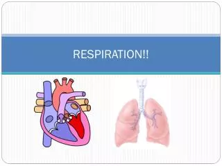
Human Respiratory System • Divided into two parts • Conducting portion, a series of passageways that carry air into and out of the gas-exchange portion of the respiratory system • Gas-exchange portion, where gases are exchanged with the blood in tiny sacs within the lungs
Conducting Portion • Carries air to the lungsand contains the apparatus that makes speaking possible • Air enters through the nose or mouth and passes through the nasal or oral cavity into the pharynx, travels to the larynx, or “voice box,” • The opening to the larynx is guarded by the epiglottis, a flap of tissue supported by cartilage which prevents food from entering the larynx when swallowing • During normal breathing, the epiglottis is tilted upward, allowing air to flow into the larynx • During swallowing, the epiglottis folds downward and covers the larynx, directing substances into the esophagus
The Human Respiratory System bronchiole pulmonary arteriole nasal cavity pulmonary venule pharynx epiglottis oral cavity larynx esophagus trachea rings of cartilage bronchioles bronchi pulmonary veins diaphragm capillary network pulmonary artery alveoli (a) Human respiratory system (b) Alveoli with capillaries
Don’t inhale and swallow at the same time. • The Heimlich maneuver • If an individual inhales and swallows at the same time, food can become lodged in the larynx, blocking air from entering the lungs • The use of the Heimlich maneuver clears the obstruction
Making Sound • Within the larynx are the vocal cords, bands of elastic tissue controlled by muscles • Muscular contractions cause the vocal cords to partially obstruct air passage through the larynx • Exhaled air causes the vocal cords to vibrate, producing the tones of speech or song • Stretching the cords changes the pitch of the tones, which can be articulated into words by movements of the tongue and lips
Human Respiration Structure • Inhaled air travels past the larynx into the trachea, a flexible tube with walls are reinforced with semicircular bands of stiff cartilage • Trachea splits into 2 bronchi, one leading to each lung • Inside the lung, each bronchus branches repeatedly into smaller tubes called bronchioles • Bronchioles lead to microscopic alveoli, tiny air sacs where gas exchange occurs
Gas Exchange • Occurs in the alveoli • Alveoli cluster at the end of each bronchioles (think:grapes) , providing 1,500 square feet of surface area for diffusion • A network of capillaries covers the alveolar surface • The walls of the alveoli consist of a single thin layer of epithelial cells • The respiratory membrane, through which gases diffuse, consists of epithelial cells of the alveoli and the endothelial cells that form the wall of the capillary, across which gas exchange occurs
Alveoli • Well adapted for gas exchange • Alveolar walls and capillary walls are only one cell thick, gases diffuse a short distance to move between the environment and blood • Alveoli are coated with a thin layer of watery fluid containing surfactant, which prevents the alveolar surfaces from sticking together and collapsing when air is exhaled • Gases dissolve in this fluid as they pass in and out of the alveolar air • Surfactant - compounds that lower the surface tension
Gas Exchange Between Alveoli and Capillaries to the pulmonary vein from the pulmonary artery capillary alveolar membrane respiratory membrane (air) CO2 O2 surfactant fluid Oxygen diffuses into the red blood cells Carbon dioxide diffuses into the alveolus
How are O2 and CO2 Transported? • Oxygen and carbon dioxide are transported in blood using different mechanisms • Blood picks up oxygen from the air in the lungs and supplies it to the body tissues, simultaneously absorbing CO2 from the tissues and releasing it into the lungs • These exchanges occur because diffusion gradients favor them • In the lungs, O2 is high and CO2 is low, whereas in body cells, CO2 is high and O2 is low
Oxygen Transport • 90% of O2 carried by the blood, bound to hemoglobin • Each hemoglobin molecule can carry up to four O2 molecules, each bound to one of four iron-containing heme groups • As oxygen binds hemoglobin, the protein changes its shape, which alters its color • Oxygenated blood is bright cherry-red • Deoxygenated blood is maroon-red
Oxygen Transport surfactant fluid (air in alveolus) respiratory membrane red blood cells O2 alveolar wall O2 hemoglobin cells of body tissues (plasma) capillary walls O2 (extracellular fluid) (a) O2 transport from the lungs to the tissues
Carbon Dioxide Transport • CO2 from cellular respiration in the body cells diffuses into nearby capillaries, then is carried in the bloodstream to the respiratory membranes of the alveoli • Alveolar capillaries have a higher CO2 concentration than that of the alveolar air • Thus, CO2 diffuses down a concentration gradient into the alveolar air, which is exhaled
Carbon Dioxide is Transported 3 Ways • As bicarbonate ions (70%) • Bound to hemoglobin (20%) • Dissolved in plasma as CO2 (10%) • Bicarbonate ions (HCO3–) are formed in red blood cells when CO2 combines with water, using the enzyme carbonic anhydrase • CO2 + H2O CO2 + HCO3–
The reaction producing bicarbonate ions is reversed as the blood flows through capillaries surrounding the alveoli, where CO2 is low: • H+ + HCO3– CO2 + H2O • As CO2 leaves the blood and diffuses into the alveoli, it diffuses back into red blood cells, where it recombines with H+, regenerating CO2 and H2O • The CO2 then diffuses into the air in the alveoli, which is exhaled from the lungs while the H2O remains in the blood
Carbon Dioxide Transport 1 CO2 CO2 2 CO2 CO2 CO2 CO2 3 H2O CO2 HCO3– + H+ 5 HCO3– CO2 CO2 + H2O H+ HCO3– 4 CO2 (b) CO2 transport from the tissues to the lungs
Inhalation, Exhalation • Air is inhaled actively and exhaled passively • Breathing occurs in two stages • Inhalation, when air is drawn into the lungs • Exhalation, when air is expelled from the lungs • Inhalation occurs when the chest cavity is enlarged • The lower boundary of the chest cavity is formed by the diaphragm, which domes upward when relaxed • During inhalation, the diaphragm is contracted, which pulls it downward, and the rib muscles contract, lifting the ribs up and outward
Exhalation • Exhalation occurs spontaneously, when the muscles that cause inhalation are relaxed • As the diaphragm relaxes, it domes upward; at the same time, the ribs fall down and inward • These movements decrease the size of the chest cavity and force air out of the lungs
The Mechanics of Breathing Air moves in Air moves out Rib cage contracts Rib cage expands Lungs compress Lungs expand Diaphragm relaxes upward Diaphragm contracts downward (a) Inhalation (b) Exhalation
Breathing Rate • Controlled by the respiratory center of the brain • Located in the medulla portion of the brain, just above the spinal cord • Nerve cells in the respiratory center generate cyclic action potentials that cause contractions (followed by passive relaxation) of respiratory muscles • The respiratory center receives input from sources and adjusts the breathing rate and volume to meet the body’s needs • Primarily modified by CO2 receptors located in the medulla that adjust the breathing rate to maintain a constant low level of CO2 in the blood, while also ensuring that O2 levels remain adequate • As a backup system, there are also O2 receptors in the aorta and carotid arteries that stimulate the respiratory center to increase the rate and depth of breathing if O2 levels in the blood drop