Download
1 / 42
430 likes | 729 Views
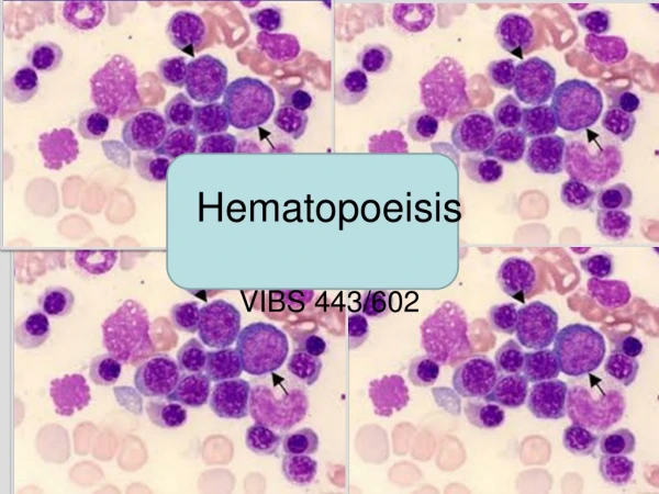
hematopoeisis. Bone marow examination Normal bone marrow 1. Haemopoietic cells. Morphology of haemopoietic cells Granulocytes & their precursors – 60% Erythroid precursors – 20 % Lymphocytes & Monocytes & their precursors – 10% Unidentified or disintegrating cells – 10%

E N D
Bone marow examination Normal bone marrow 1. Haemopoietic cells. Morphology of haemopoietic cells • Granulocytes & their precursors – 60% • Erythroid precursors – 20 % • Lymphocytes & Monocytes & their precursors – 10% • Unidentified or disintegrating cells – 10% 2. Myeloid : erythroid ratio – 3-4 :1
Anaemia • Anaemia may be defined as a state in which the blood Hg is below the normal range fro the age & sex of the patient Normal count/Range Hemoglobin • Male – 13 –18 gm/dl • Female -11.5 – 16.5 gm/dl Red cell count – • Male - 4.5–6.5 million/cu mm of blood • Female – 3.8–4.8 • Reticulocyte count - 0.2–2.0%
Factors necessary for Erythropoiesis. A] General Factors • 1] Diet-Protein is essential for synthesis of Globin in of Haemoglobin • Iron-essential for heam production • Others-Cu, Mg, Cobalt, Ca etc. • 2] Hypoxia-hypoxia causes Liberation of hormone Erythropoietin which stimulate bone marrow for Erythropoiesis • 3] Erythropoietin • 4] Endocrine gland- Adrenal Gland, Pituitary Gland, Thyroid Gland. B] Maturation factors • 1] Vit. B12 • 2] Folic Acid • 3] Intrinsic Factor of Castle-It helps in absorption of Vit. B12 from gut and thus indirectly help in maturation.
Classification of Anemia Morphological & Aetiological Classification Morphological classification • Based mainly on MCV & MCHC. MCH may be included 1.Microcytic Hypochromic aneamia • MCV, MCHC & MCH are below normal • E.g. – IDA, thalassaemia, anaemia of chronic diseases
2. Macrocytic anaemia • MCV is above normal . MCHC is normal • E.g. megaloblasticanaemia 3. Normocytic Normochromic anaemia • MCV, MCHC MCH are within normal range • E.g. acute blood loss, haemolytic anemia
Aetiological Classification This classification is based on pathophysiology & cause • Impaired RBC production • Hemolytic anemia • Blood Loss
Impaired RBC production A. Deficiency of essential nutrients (Deficiency anaemia) • Iron deficiency – most common cause • Vitamin B12 deficiency • Folic acid deficiency • Combined deficiency Others – protein calorie malnutrition , vitamin C deficiency
B. Depression of erythropoiesis • Anemia of chronic disease – Chronic renal failure . Liver diseases, Malignancy • Invasion of bone marrow – leukaemia , secondary carcinoma • Aplastic anemia
B. Haemolytic anemia • Intracorpuscular defect • Extracorpuscular defect C. Blood Loss • Acute Blood loss – loss of large volume of blood over a short period • Chronic blood loss e.g hookworm infestation, bleeding peptic ulcer, piles, menorrhagia
Clinical features of anemia • Tiredness , Fatigue • Lethargy • Palpitaion • Pallor is the most important physical sign
Laboratory diagnosis of anemia • Hb estimation • Diagnosis of Morphological type of anaemia • Diagnosis of aetiological type of anemia – discussed under individual type of anemia
Packed cell volume (PCV) • Is the volume of red cells in relation to that of whole blood PCV = MCV X red cell count Normal values • 40 – 45 % male • 37- 47 % female MCV (Mean corpuscular volumes ) • Indicates the average volume red cells • Normal 76 - 96 fl (femolitre)
MCH (mean corpuscular hemoglobin ) • Indicates average weight of Hb contained in each cell • Does not take into count cell size • Obtained by dividing Hb by red cell /liter & multiplied by 1013 If Hb 15g/dl Red cell – 5X10¹²/L MCH = 15X1013 / 5X1012 = 30 pg • Normal 27-32 pg
MCHC (mean corpuscular haemoglobin concentration) • Indicates the average concentration of Hb within average red cells • Usually 3 times higher that the whole blood Hb conc. • Obtained by dividing Hb by PCV /liter If Hb 15g/dl PCV – 0.45l/l MCHC = 15/0.45 = 33.3g/dl • Normal – 31-35 g/dl
Diagnosis of morphological type of anemia 1. Examination of peripheral blood film 2. Determination of Red cell absolute value • MCV & MCHC are below normal values – microcytic hypochromic anemia • MCV is above normal & MCHC is normal – macrocytic anemia • MCV & MCHC are normal – normocytic normochromic aneamia
Iron deficiency anemia Most common type of anemia Total body iron content • Male – upto 6gm • Female – 2gm 80% of functional body iron is found in hemoglobin, rest found in myoglobin and iron-containing enzymes (e.g.catalase & cytochromes). The iron storage pool, represented by hemosiderin and ferritin-bound iron (15% to 20% of total body iron.) • Stored iron is found mainly in the liver, spleen, bone marrow, and skeletal muscle. Male Fem
Absorption • Most absorbed in Duodenum
Causes /pathogenesi of iron deficiency 1. Inadequate iron intake • Nutritional deficiency – deficient diet 2. Impaired absorbtion – coeliac disease, tropical sprue, gastrectomy or gastro-enterostomy
Causes /pathogenesi of iron deficiency 3. Increased physiological demand • During period of growth in children • During reproductive life in female – menstruation, pregnancy, parturation & lactation increase the physiological requirement fro iron 4.Chronic blood loss – the gastrointestinal tract (e.g., peptic ulcers, colonic cancer, hemorrhoids, hookworm disease) or the female genital tract (e.g., menorrhagia, cancers)
Laboratory diagnosis of IDA Investigation & findings 1. Blood Picture • Hb – Variably reduced • Blood film – hypochromic, microcytic, anisocytosis. Poikilocytosisin severe cases target, elliptical, oval & pencil cells • Haematocrit – reduced • MCV – reduced(microcytosis) MCHC – reduced (hypochromic) & MCH is reduced
2. biochemical findings (confirm IDA) • Serum iron - reduced • Serum ferritin - reduced • Total iron binding capacity – increased • Percentage saturation of iron binding protein – decreased 3. Bone marrow • Although erythropoietin levels are increased, the marrow response is blunted by the iron deficiency, and thus the marrow cellularity is usually only slightly increased Diagnostic criteria include • Anemia, hypochromic and microcytic red cell indices, low serum ferritin and serum iron levels, low transferrin saturation, increased total iron-binding capacity, and, ultimately, response to iron therapy
4. Further Investigation • Stool examination for • Ova of hookworm & • Occult blood • Urine examination for haematuria • Other investigations depending on clinical findings
Megaloblastic aneamia • MA are characterized by formation of morphologically abnormal (enlarged) nucleated red cell precursors called megaloblasts in the bone marrow. • The change occurs due to deficiency of vitamin B12 or folate
Vitamin B12 • Abundant in all animal foods, including eggs and dairy products, and is resistant to cooking and boiling. • Even bacterial contamination of water and non animal foods can provide adequate amounts. • It is stored in the liver, which normally contains reserves that are sufficient to support bodily needs for 5 to 20 years • As a result, deficiencies due to diet are rare and are virtually confined to strict vegans
Metabolism of vitamin B12 • Peptic digestion release dietary vitamin B12, • Binds to salivary B12-binding proteins called R binders. • R-B12 complexes transported to duodenum • Processed by pancreatic proteases to releases B12, • Attaches to intrinsic factor (gastric juice) • The intrinsic factor-B12 complex passes to the distal ileum • Attaches to the epithelial intrinsic factor receptors - absorption of vitamin B12. • B12 bound to transport proteins called transcobalamins, which then deliver it to the liver and other cells of the body.
Causes of Megaloblastic Anemia Vitamin B12 Deficiency • Decreased intake • Inadequate diet, vegetarianism • Impaired absorption • Intrinsic factor deficiency - Pernicious anemia, Gastrectomy • Malabsorption states • Diffuse intestinal disease e.g. lymphoma • Ileal resection, ileitis • Competitive parasitic uptake - Fish tapeworm infestation • Bacterial overgrowth in blind loops and diverticula of bowel • Increased requirement • Pregnancy, hyperthyroidism, disseminated cancer
Folate metabolism • Best sources - fresh uncooked vegetables and fruits. • The principal site of intestinal absorption is the upper third of the small intestine • Conversion from dihydrofolate to tetrahydrofolate by the enzyme dihydrofolatereductase is particularly important. • Tetrahydrofolate involved in the synthesis of purines and thymidylate, the building blocks of DNA,
Megaloblastic anemia Folic Acid Deficiency • Decreased intake • Inadequate diet—alcoholism, infancy • Impaired absorption • Malabsorption states • Intrinsic intestinal disease • Increased metabolism • Anticonvulsants, oral contraceptives • Increased loss • Hemodialysis • Increased requirement • Pregnancy, infancy, disseminated cancer, markedly increased hematopoiesis • Impaired use • Folic acid antagonists – e.g.Methotrxate
Pathogenesis of Megaloblastic anemia • Deficiency of Vit B12/folic acid • Impairment of DNA synthesis, • Results in a delay in nuclear maturation and cell division. • Synthesis of RNA and cytoplasmic elements proceeds at a normal rate • Hematopoietic precursors show nuclear-cytoplasmic asynchrony • Undergo apoptosis in the marrow (ineffective hematopoiesis)
Laboratory diagnosis of MA Blood • Hb– reduced • Blood film • RBC- many oval macrocytes, Anisocytosis, poikilocytosis, Polychromatic & stippled cells, howell-jolly bodies • White cells – hypersegmented neutrophils are always present (4-9 lobes) hypersegmented neutrophil with a six-lobed nucleus Megaloblastic blood compared to normal blood
Haematocrit (PCV)– reduced • Red cell count – reduced • MCV is high, MCH is high, MCHC is normal • WBC – leucopenia (neutropenia) • Platelets – thrombocytopenia • Reticulocyte count – increased 2. Biochemical findings • Serum iron & ferritin – increased • Serum bilirubin – may be slightly increased in Vit B12 deficiency • Lactate dehydrogenase – increased Male Fem
3. Bone marrow • Cellularity – Hypercellular (increased numbers of megaloblasts) • M:E ratio is reduced or even reversed • Erythropoiesis - Megaloblastic erythropoiesis • Granulopoiesis is active – giant metamyelocyte with u shaped nucleus • Iron in large amount. Sideroblast – increased A to C, Megaloblasts-various stages of differentiation
4. Special tests for vitamin B12 deficiency • Serum vitamin B12 assay • Schilling test – detects ability of the body to absorb vit B12 after correction.Radioactive cobalt labeled vit B12 is used for the test . A small oral dose is given . Radioactivity in the urine is measured • Methylmelonic acid excretion in urine - increased • Therapeutic trail - Response to vit B12 administration
5. Special Tests for folate deficiency • Serum folate assay • Red cell folate assay • Theurapeutic trail - Response to folic acid administration
Pernicious anemia PA is a vitamin B 12 deficiency megaloblasticanaemia Pathogenesis • PA occurs due to failure of secretion of Intrinsic factor by the stomach due to permanent gastric atrophy • Vit B12 in food not absorbed (gastrectomy, resection of ileum ) Special tests • Pentagastrin fast achlohydria • Anti-intrinsic factor & anti-parietal cell antibodies in serum
Diagnosis is made by • (1) low serum vitamin B12 levels, • (2) normal or elevated serum folate levels, • (3) serum antibodies to intrinsic factor, • (4) moderate to severe megaloblastic anemia, • (5) leukopenia with hypersegmented granulocytes, and • (6) a dramatic reticulocytic response (within 2-3 days) to parenteral administration of vitamin B12.