Download
1 / 20
210 likes | 375 Views
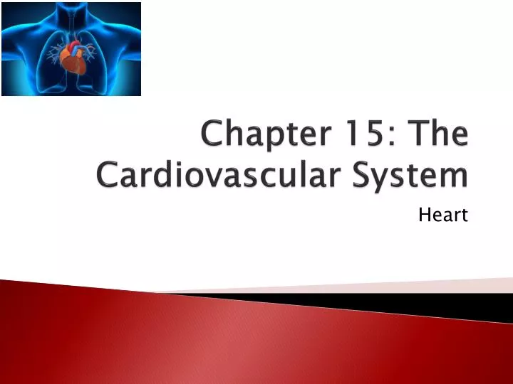
Chapter 15: The Cardiovascular System. Heart. Introduction to the Heart. Hold up your clenched fist Your heart is about the size of your fist Varies by gender, and age of the owner Age changes are due to increases in the size of cells, not number of cells

E N D
Introduction to the Heart • Hold up your clenched fist • Your heart is about the size of your fist • Varies by gender, and age of the owner • Age changes are due to increases in the size of cells, not number of cells • Average mass of a heart 230-350 grams (0.5 – 0.8 pounds)
STRUCTURE AND ORGANIZATION OF THE HEART • Location and Coverings of the Heart • Heart located between two lungs in thoracic cavity • 2/3 of mass is left of body’s midline • Apex pointed end formed at tip of left ventricle • Base formed by atria, mostly left atrium • Major blood vessels enter and exit at the base
Pericardium: membrane that surrounds and protects the heart and holds it in place • Two parts of pericardium: (1) Fibrous pericardium (2) Serous pericardium
Inflammation of the Pericardium • Pericarditis build up of pericardial fluid; compresses the heart = cardiac tamponade
Heart Wall • Three layers: • EXTERNAL (1) epicardium or visceral layer of serous pericardium • Thin, transparent outer layer of the wall • Composed of mesothelium and connective tissue
MIDDLE (2) myocardium • Bulk of heart wall • Consists of cardiac muscle • Responsible for pumping action of heart
Cardiac muscle two separate networks connected by intercalated discs • Atrial • Ventrical • Gap junctions in the discs allow action potential conduction from one muscle fiber to the next
INTERNAL (3) endocardium • Thin layer of squamous epithelium lining inside of myocardium • Covers valves of heart and tendons attached to the valves • Continuous with epithelial lining of large blood vessels
Chambers of the Heart • Four chambers: • Two upper are the atria • Right atrium • Left atrium
Two lower are the ventricles • Right ventricle • Left ventricle
Great Vessels of the Heart • Right atrium receives deoxygenated blood through three veins… • Superior vena cava • Inferior vena cava • Coronary sinus
Right ventricle pumps blood into the pulmonary trunk • Right pulmonary artery goes to right lung • Left pulmonary artery goes to left lung • Blood becomes oxygenated
5 3 3 6 6 1 4 4 7 2 8
Valves of the Heart (4) • Prevent blood from flowing backward • Composition: dense connective tissue covered by endothelium • TWO Atrioventricular (AV) valves lie between the atria and ventricles • Between the right atrium and right ventricle is the AV called the (1) tricuspid valve, which has the three cusps
Chordae tendineae • tendon-like chords that connect the pointed ends of the cusps to cardiac muscle projections on inner surface of the ventricles • prevent the cusps from pushing up into the atria when the ventricles contract
AV between the left atrium and left ventricle is called the (2) bicuspid (mitral) valve • It has two cusps
TWO Semilunar valves are located near origin of pulmonary trunk and aorta • Prevent blood from flowing back into the heart • Pulmonary valve • Aortic valve Both consists of three semi-lunar cusps attached to the artery wall