Download

1 / 13
130 likes | 179 Views
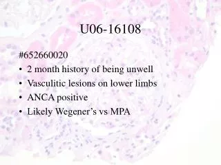
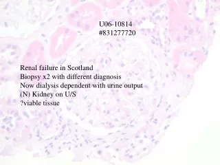
U06-10814 #831277720. Renal failure in Scotland Biopsy x2 with different diagnosis Now dialysis dependent with urine output (N) Kidney on U/S ?viable tissue. Case Summary: ES

E N D
U06-10814 #831277720 Renal failure in Scotland Biopsy x2 with different diagnosis Now dialysis dependent with urine output (N) Kidney on U/S ?viable tissue
Case Summary: ES 67 year old Caucasian female, retired school teacher and mother of 3 with significant past medical history of (1) ureteric surgery 1965; (2) G6P3 – 3 miscarriages between 2nd and 3rd child; (3) Ex-smoker – quit 20 years ago; previously smoked ½ pk/day for 10 years. HPI: Jan-March 2005: Traveled to Vietnam; developed severe diarrhea upon return to Canada; Blastocystis hominis; treated with Cipro for 1 month with resolution of diarrhea however next 6-9 months general health intermittently poor; malaise; fatigue; constitutional symptoms. Sept 2005: Traveled to Scotland to visit daughter (vet); URTI/pneumonia/treated with levoquine for 10 days; questionable interstitial fibrosis noted on CXR Dec 2005: presented to Dumfries hospital with SOBOEx/ fever/rash/wt loss
During admission in Scotland (Dec 2005 – May 2006), several findings: Hepatomegaly with transaminitis – liver biopsy – steatosis heart murmur noted; ?Q fever – treated with doxycycline x 10 d;serology negative Grp B strep isolated from throat swab – treated with amoxil for 10 d Bilateral pleural effusions and pericardial effusions - ?vasculitis started on steroids Mantoux positive but history of BCG vaccination CT scan showed mediastinal lymphadenopathy – biopsy reported on Feb 21/06 as MAC – treated with ethambutol/clarithromycin Opthamology consulted re screening pre-ethambutol – dx retinal vasculitis Temporal aa bx – negative; bone marrow biopsy negative; blood cultures negative; viral serology negative; autoantibodies negative
March 2006: tap of pleural effusion – transudate; renal function noted to be declining; clarithromycin d/c and azithromycin started; renal dosing of ethambutol; noted to have 2 g/day proteinuria; steroid increased; thrombocytopenic (80 lowest) and MAHA (90 lowest) noted; renal biopsy #1 performed in Dumfries and renal biopsy #2 performed in Glasgow. Based on biopsy #1: treated with IV solumedrol/Cyph; based on biopsy #2: plasma exchange initiated. March 17, 2006: started on hemodialysis; rapid afib – started digoxin Rheumatology opinion: not scleroderma; MAC treatment d/c. May 1. 2006: 2nd pleural effusion tap: transudate. Transferred to UAH May 19, 2006 (treated with 10 weeks of steroids at this point). Issues while at UAH:
Renal failure – dialysis dependent; producing ~ 400-600 cc/d with large doses of diuretics; MRI/MRA kidneys – normal size kidneys; extrarenal pelvis on Rt; all serology repeated and negative including ANCA, ANA antiphospholipid Ab and Anti SCL70, complements normal; June 1st renal biopsy. Viral serology repeated negative. Steroids being tapered. Pulmonary status – new airspace disease LLL; persistent L pleural effusion>R; CT chest – cardiomegaly; bilat pl effusions; LUL and LL ground glass opacification persistent; past granulomatous infection; echocardiogram June 14: no pericardial effusion; good wall motion; Lt pl effusion; bronchoscopy with BAL – negative; Bone marrow repeated June 2nd: negative culture; all antibiotics stopped; acute deterioration with hypoxia June 16th – no worsening of Lt pl effusion/ MAC a concern/ started on Pip/Tazo/Azith/Septra– U/s guided tap June 22 Neurologic status – CT head May 31 – small vessel ischemic changes; CT head for acute deterioration June 21 no change Persistent normocytic anemia; platelet count normal/high; MRSA nose and groin positive initially; now bacteremic from CVC; on vanco Depression – on Celexa; psychiatry following
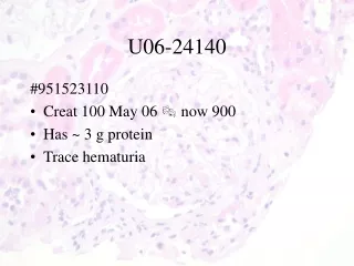
IF • IgG-negative • IgA- negative • IgM- mild mesangial staining • C3- moderate peritubular capillary staining • C1q-negative • Kappa-negative • Lambda-negative • Fibrin- moderate interstitial staining • Albumin- negative
IgM IgM
Diagnosis: Renal Biopsy: Thrombotic Microangiopathy in a relatively quiescent phase with few obvious thrombi.