Download
1 / 59
650 likes | 898 Views
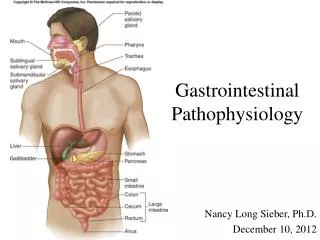
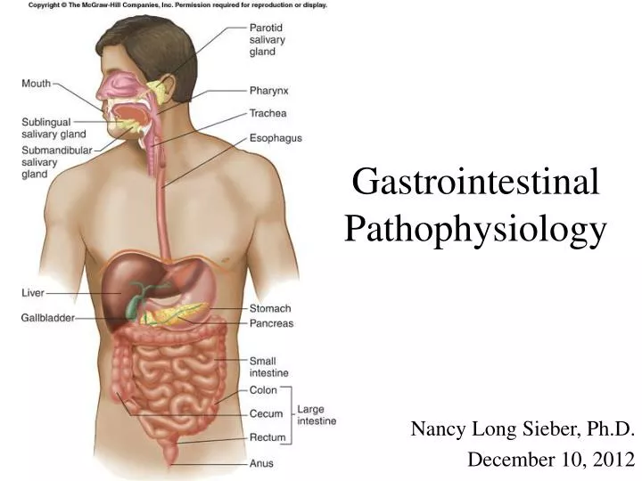
Gastrointestinal Pathophysiology. Nancy Long Sieber, Ph.D. December 10, 2012. A Guided Tour of the GI Tract with pathological detours. Mouth and Pharynx Chewing and Swallowing. Swallowing. Swallowing requires f unctional teeth to

E N D
Gastrointestinal Pathophysiology Nancy Long Sieber, Ph.D. December 10, 2012
Swallowing Swallowing requires functional teeth to break down food, salivary glands to soften and lubricate it and neural coordination of both skeletal and smooth muscle. What can go wrong?
What can interfere with swallowing? • Dental illness can interfere with chewing • Certain illnesses and some medications interfere with salivary secretion. • Sjogren’ssyndrome (autoimmune attack on salivary glands) interferes with the production of saliva, making it more difficult to chew and swallow food. • Many medications have dry mouth as a side effect. Eg: parasympathetic antagonists such as atropine (in anti-diarrhea drugs), and sympathetic agonists (in decongestants) • Certain CNS diseases and conditions can interfere with swallowing, such as amyotrophic lateral sclerosis (ALS), spinal cord injury and stroke.
The EsophagusWhat if what you swallow doesn’t stay swallowed?
Factors that increase the likelihood of Gastroesophageal Reflux • Being an infant • Obesity • Pregnancy • Postural changes (bending over, lying down) • Smoking • Certain foods relax the LES – why are these popular to have after dinner? • Peppermint • Coffee • Alcohol • People with lupus have more problems with GERD due to connective tissue problems
Hiatus Hernia http://www.merck.com/media/mmhe2/figures/fg123_1.gif
Barrett’s Esophagus http://www.barrettsinfo.com/figures/fig3a_4.jpg
The StomachMechanical mixing with acid, some enzymatic digestion.
The stomach is protected from acid by a layer of mucus and bicarbonate http://en.wikibooks.org/wiki/Medical_Physiology/Gastrointestinal_Physiology/Secretions
Helobacter pylori passes though mucus layer that protects the stomach. Contact with stomach acid keeps the mucin lining the epithelial cell layer in a spongy gel-like state. This consistency is impermeable to H pylori. However, the bacterium releases urease which neutralizes the stomach acid. This causes the mucin to liquefy, and the bacterium can swim right through it. http://www.nsf.gov/news/news_images.jsp?cntn_id=115409&org=NSF
NSAIDS interfere with prostaglandin synthesis, which blocks production of prostaglandin. This blocks production of mucus and bicarbonate, and increases acid production, making the stomach vulnerable to injury from acid and enzymes. http://www.gastrosource.com/11674565?itemId=11674565
Bariatric Surgery Currently used for severely obese people who have not been able to lose weight through diet and exercise. Gastric banding was recently approved for less seriously obese people (eg: a 5’5” person who weighs 180 lbs)
Carbohydrate Digestion http://media.tiscali.co.uk/images/feeds/hutchinson/ency/thumbs/0013n025.jpg
Protein Digestion http://media.tiscali.co.uk/images/feeds/hutchinson/ency/thumbs/0013n023.jpg
Fat Digestion http://media.tiscali.co.uk/images/feeds/hutchinson/ency/thumbs/0013n024.jpg
GI fluid imbalance can be caused by: • Vomiting – forceful emptying of stomach and upper intestinal contents through the mouth • Diarrhea – increased frequency of defecation and volume of feces. Diarrhea is the leading cause of death among children in the developing world
Consequences of prolonged vomiting • Dehydration • Metabolic Alkalosis (due to loss of gastric acid) • Low serum K+ (due to action of aldosterone, which is released after prolonged dehydration). • Malnutrition
Diarrhea • Potential for massive fluid loss, leading to hypotension. • Can cause acidosis, since intestinal contents are alkaline. • Can cause hypokalemia (low K+) since intestinal contents are high in K+.
Osmotic Diarrhea:Lactose intolerance results from a lactase insufficiency. Undigested lactose remains in the intestine, and osmotically draws water into the intestine, causing diarrhea. http://www.food-info.net/images/lactase.jpg
Secretory Diarrhea: Cholera. Cholera toxin increases the intracellular levels of cAMP. This leads to an increase in chloride secretion into the small intestine. Water follows osmotically http://www.surrey.ac.uk/SBMS/ACADEMICS_homepage/mcfadden_johnjoe/img/Cholera%20toxin.jpg
Celiac Disease • Results from intolerance to gluten, specifically the gluten breakdown product gliaden. • Considered to be an autoimmune disease – the person makes antibodies against gliadin, which cross react with proteins in the small intestine. • This causes inflammation and damage to the intestinal villi.
Celiac disease: Effect on cells in the small intestine These images show cells collected during a small bowel biopsy.The slide on the left shows normal intestine and the finger-like villi, which absorb nutrients from food. The slide on the right, by contrast, shows the effects of celiac disease. The villi can no longer absorb nutrients because the intestine is inflamed and swollen. http://www.riversideonline.com/health_reference/Articles/DG00023.cfm
Consequences of celiac disease • Pain and diarrhea, inflammation of intestinal villi. • Malabsorption of nutrients leading to • Osteoporosis (from lack of calcium) • Anemia (from lack of iron) • Short stature, when it develops in childhood (from general malnutrition) • Miscarriage, neural tube defects (lack of folic acid, and other nutrients)
Hemorrhagic Diarrhea • Infectious diarrhea leading to blood in stools • Example: recent outbreaks of E. coli 0157:H7 • May be a consequence of factory farming practices • Grows well in corn-fed factory farmed beef • Runoff contaminates vegetable farms
Diarrhea due to altered motilityDumping Syndrome • Typically caused by gastric surgery for peptic ulcer disease, or by gastric by-pass surgery. Part of the stomach typically including the pyloric sphincter is removed , and the meal instead of very slowly entering the duodenum can enter very quickly. • The increased osmolarity of the contents results in very rapid movement of water into the intestines. • In addition, the very rapid presentation of carbohydrates to the intestinal mucosa and their absorption causes a big spike in blood sugar resulting in a large increase in insulin.
Important GI Organs:The Pancreas and the Liver And, of course, What can go wrong with them?
The pancreas releases the major digestive enzymes, which are needed to break down starch, proteins and fats.
Pancreatic enzymes are usually released in an inactive form.
Acute Pancreatitis • Results from acute inflammation and autodigestion of pancreas • Causes • Alcohol abuse – activates trypsinintracellularly, also can cause inflammation of the sphincter of Oddi & reduce its tone • Gallstones – can be impacted in the pancreatic duct.
Manifestations of acute pancreatitis: • Pain • Nausea & vomiting • Fever • Shock • Amylase and lipase in blood • Jaundice • Acidosis • Hyperkalemia
Chronic Pancreatitis • Characterized by fibrosis and calcification • Loss of exocrine function • Usually caused by alcoholism
Pancreatic Insufficiency • Usually caused by chronic pancreatitis • Leads to : • Malabsorption • Fat in stool (fat not absorbed) • Diarrhea (but not as bad as from intestinal malabsorption, such as celiac disease) • Can give replacement enzymes
Pancreatic Cancer • High mortality rate (95%) • Symptoms usually do not develop until disease has spread.
The liver does many things: • Fat digestion – via production of bile • Nutrient storage, synthesis, and mobilization • Storage of carbohydrates as glycogen and gluconeogenesis • Fat metabolism and cholesterol synthesis • Vitamin and trace mineral storage • Biotransformation of drugs and toxins • Metabolism of hormones • Production of plasma proteins • Immune surveillance
The openings (fenestrations) in the capillaries mean that the hepatocytes are essentially in direct contact with the blood. http://www.siumed.edu/~dking2/erg/liver.htm
Bile Pigments • Bile pigments are breakdown products of hemoglobin. Most important is bilirubin. • The pigments are carried to the liver, where they are combined with bile, and are converted into a form that the body can excrete. • When this process fails, and bilirubin accumulates in the tissues, it causes the skin and eyes to become yellowish.