Download
1 / 32
350 likes | 692 Views
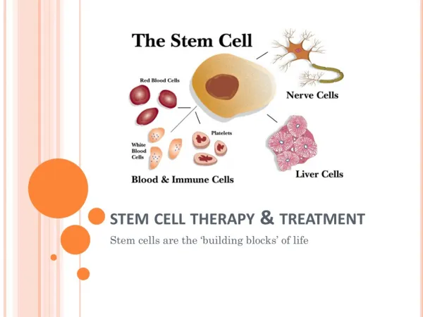
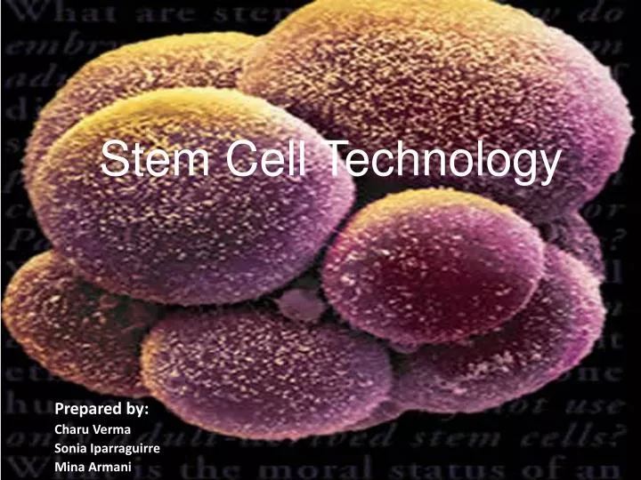
Stem Cell Technology. Prepared by: Charu Verma Sonia Iparraguirre Mina Armani. Our Research Experience. Looking at De Coppi’s method taking amniotic fluid-derived stem cells can be differentiated into all 3 germ layers, including nerve cells.

E N D
Stem Cell Technology Prepared by: Charu Verma Sonia Iparraguirre Mina Armani
Our Research Experience • Looking at De Coppi’s method taking amniotic fluid-derived stem cells can be differentiated into all 3 germ layers, including nerve cells. • Our research group decided to take this method and apply it to frozen human fibroblast that varied in age, from amniotic to 96 years old. • These methods worked, however for nerve cells it took several weeks to differentiate.
Nerve and Adipose Cell differentiation • Cells that were brought from Coriell Institute in Camden, NJ • Cells were left in incubation of 37°C at 5% CO2 for approximately 3 days till cells were confluent • Once cells were confluent, these cells were emptied and washed in HANKS media • Trypsin was then added, so cells would come off from the bottom of the flask to then be transferred to a new flask • Cells were calculated through the hemocytometer to find out how many cells per ml are in a flask, each new flask must contain at least 75,000 cells for seeding. • The amount of cells calculated were then transferred into 25cm flasks filled with Media 1, which was grown for several days until they were confluent. • Cells were confluent and passed at least 3 passages, using the same protocol as above, these cells would then go through trypsin again and then transferred into a new flask with the either nerve media or adipose media for differentiation.
Hepatic Cell Differentiation • Cells were seeded at a density 5,000 cells/cm2 along with regular Media 1 in petri dishes, coated with Matrigel, for 3 days. • The Media 1 was then changed to Hepatic media and maintained for 2 weeks • Cells were washed in HANKS and then trypsinize to be plated in collagen sandwich gels. • Cultures were maintained for up to a total of 45 days .
Muscle Cell Differentiation • Cells were seeded at a density of 3,000 cells/cm2 along with regular Media 1 petri dishes, coated with Matrigel. • After 12 hours , the media was changed to Muscle media and was then incubated for 24 hours • Incubation of cells growth were completed with the use of Muscle Media, without the ingredient 5-azaC • Changes of the Muscle Media, without 5-azaC, was done every 3 days
Nerve Cell Differentiation without DMSO • There were 6 different medias formed, 3 medias that had DMSO and 3 medias that lacked DMSO, with different nutrients in a each media. • The chamber slides that had media with all nutrients but with no DMSO were able to differentiate into nerve cells within 7 days. • Micro array was done which showed gene expression that were 35 up-regulated and 140 down regulated genes at two fold between undifferentiated and differentiated nerve cells. • Most cells were all up-regulated genes with unknown functions
Stains • Cells were emptied and wash 3 times with PBS and then fixed with 4% paraformalhyde • Differentiated nerve cells were stained positive with Glial Fibrillary Acidic Protein (GFAP ) and Neural Filament-M (NFM). • Neural Filament-M is used to stain and identify neurons. • Glial Fibrillary Acidic Protein (GFAP) is used to identify neural stem cells, astrocytes, non myelinating Schwann cells in peripheral nerves, and peripheral ganglia. • For negative controls, cells were grown in regular aMEM to see if there was immunofluorescence, which there was no any fluorescence.
Phase Contrast picture of a dividing and a dead fibroblast cell.
Human fibroblast stem cells were obtained from individuals of different age categories. They were cultured in special medium before turning them Into specialized human cells.
Special growth factors were added into the culture media to induce Differentiation of fibroblast stem cells into the adipose cells.
Different growth factors were used to induce differentiation of fibroblast stem cells into muscle cells.
A C D B Nerve cells stained with GFAP (Glial Fibrillary acidic Protein). Pictures A & B are viewed under immunoflourescent microscope. Pictures C & D are viewed under phase contrast microscope.
PLANT STEM CELL TECHNOLOGY GRADE LEVEL: 9-12 NJCCCS: 5.1, 5.2, 5.3,5.4,5.5 Lesson Objective: Student will be able to • Understand the properties and functions of stem cells. • Apply the usage of stem cells in life. • Breeding a new plant from plant somatic tissue. • Investigating the effect of plant hormones on plant cell differentiation.
Assessment • What do you know of stem cells? • Do you think plants have stem cells, and if so, where do you find them? • What is cell differentiation?
Instructional Input and Information Will be delivered using a PowerPoint presentation.
Modeling Maintaining aseptic conditions in plant tissue culture
BACKGROUND INFORMATION • Angiosperms or flowering plants are classified into two groups; monocots, and dicots. • The seeds of flowering plants consist of the cotyledon(s) and the embryo. • Monocots are the seeds with one cotyledon and dicots are the seeds with two cotyledons. • By removing the embryo from the cotyledons and then placing the embryos on agar having varying hormone concentrations, one can see the influence that the hormones have on the growth of the embryo.
The embryo is composed of the epicotyl which is the portion of the seed above the attachment of the cotyledons and gives rise to the stem and leaves. • The hypocotyl is the portion of the stem below the attachment of the cotyledons. The part of the embryo that contains the root apical meristem becomes the first primary root of the seedling. • Different parts of the embryo may react differently to either the type of hormone or the concentration of the hormone.
SAFETY CONSIDERATIONS 1. Students should use goggles and aprons when handling the bleach and alcohol solutions. 2. The open flame of the Bunsen burner should be a safe distance from the alcohol when using the flame technique of sterilization. If no Bunsen burner is available, one can use a beaker filled with glass beads on a hot plate at 250 C to disinfect the tools. 3. Students must be cautioned about the sharp edges that are found on scalpels. 4. When doing tissue cultures, the work area and equipment should be kept as sterile as possible to prevent contamination from molds or other microorganisms.
Material • (based on a class of 24 students): • Sterile beaker (one per group, used for surface sterilization of seeds) • Sterile scalpel (one per group) • Sterile forceps, needle nose (two per group) • Bunsen burner (one per group) • Sterile Petri dishes or baby food jars that can be sterilized with media in them (number will vary depending on the teacher's needs) • Lima bean seeds, corn seeds and other seeds of the teacher's choice • REAGENTS • (based on a class of 24 students) • 70% ethanol, in a bottle (0.5 L/class) • 95% ethanol, in a jar with a lid • 10% bleach solution, in a bottle(0.5 L/class) • 50% bleach solution, in a squirt bottle (1.0 L) • Sterile distilled water, in container for each station (1.0 - 1.5 L/class) • Murashige and Skoog basal medium with sucrose and agar (Sigma M9274) • Indoleacetic acid, 0.3 g/L of medium • Kinetin, 1 mg/L of medium • Adenine, 80 mg/L of medium • **Teachers have to prepare the solutions and media aseptically prior to the actual activity. This would take approximately 2 hours.
PROCEDURE DAY 1 • Each group will use the lima beans to represent dicots, and corn to represent monocots. • Students will put at least 15 seeds of each type in a beaker. They will submerge the seeds in water, cover with aluminum foil, and allow all seeds to soak for 24 hours. DAY 2 • Maintaining sterile conditions following the given instructions, students will separate the embryos from the seeds. • Students will grow the embryos by placing them on provided sterile media containing different concentrations of hormones. DAYS 3 – 7 • Students will observe and record any changes in the developing embryos that are growing on the culture media for at least one week. They will record the results and sketch the embryo indicating where most of the growth took place.
Guided Practice/Monitor and Adjust Day 1 Assignment: • Students should investigate different parts of a plant embryo to discuss in class the next day. • Teacher should check for students understanding and guide them through if needed. Day 2 Assignment: • Students should explore the function of Kinetin, IAA, and Adenine on the growth of plant embryo. • Teacher should check for students understanding and guide them through if needed. Day 3 Assignment: • Students should hypothesize the effect of the usage of different ratio of Kinetin to IAA on embryonic plant growth. • Assessment of Day 3 will be done at the end of the activity.
Critical QUESTIONS Answer in complete sentences. 1. Describe the pattern of growth on the different dishes. 2. On which of the dishes did most of the growth take place? Explain the relationship between hormone concentration and growth of the embryos. 3. Explain any differences between the growth of the epicotyl and the hypocotyl. 4. If the embryos were still attached to the cotyledons, what would have been the primary source of energy for the developing plant? 5. How would you relate your experiment to stem cell growth and differentiation?
Closure • Teacher will give an overview of the lab and the concept under investigation. • Teacher will encourage a class discussion on the student’s hypothesis and the results they observed. • Students will write a lab report based on the given protocol, following the attached rubric.
Acknowledgement Special thanks to the following for providing us with the opportunity to participate in the PARSE Institute Workshop, Summer of 2009. Dr. James Clayton Director of Educational Programs of the PARSE Institute Dr. Jose L. Lopez Director of Scientific Program, PARSE Institute Dr. Leonard Sciorra Department Chairman of Applied Science and Technology John Ruppert Adjunct Professor SPC Biology Department
Finally Thanks to all of the PARSE Fellows for sharing their ideas and support during this workshop.