Download
1 / 28
350 likes | 661 Views
T-61.181 Biomedical Signal Processing. ECG Signal Delineation And Compression. Chapters 6.2.6 – 6.3 18th November. Outline. ECG signal delineation Definition (What) Clinical and biophysical background (Why) Delineation as a signal processing (How) ECG signal compression

E N D
T-61.181 Biomedical Signal Processing ECG Signal Delineation And Compression Chapters 6.2.6 – 6.3 18th November
Outline • ECG signal delineation • Definition (What) • Clinical and biophysical background (Why) • Delineation as a signal processing (How) • ECG signal compression • General approach to data compression • ECG signal compression (Intrabeat/Interbeat/Interlead) • Summary
Part I. EGC signal delineation
Delineation - Overview • Aim – Automatically decide/find onsets and offsets for every wave (P, QRS, and T) from ECG signal (PQRST-complex) • Note! Experts (Cardiologist) use manual/visual approach
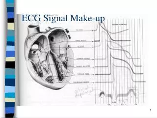
Why? • Why – Clinically relevant parameters such as time intervals between waves, duration of each wave or composite wave forms, peak amplitudes etc. can be derived • To understand this look how ECG signal is generated
What Are We Measuring? • ECG gives (clinical) information from generation and propagation of electric signals in the heart. • Abnormalities related to generation (arrhythmia) and propagation (ischemia, infarct etc.) can be seen in ECG-signal • Also localization of abnormality is possible (12 lead systems and BSM)
QRS duration Bundle brand block depolarization • ST segment ischemia • QT interval ventricular fibrillation • PR interval SA ventricles Clinically Relevant Parameters
Signal Processing Approach to Delineation (How) • Clinical importance should now be clear • Delineation can also be done manually by experts (cardiologist) expensive and time consuming. We want to do delineation automatically (signal processing) • No analytical solution performance has to be evaluated with annotated databases
Building Onset/Offset Detector • Many algorithms simulate cardiologist manual delineation (ground truth) process: • Experts look 1) where the slope reduce to flat line 2) respect maximum upward, downward slope • Simulate this: define the boundary according to relative slope reduction with respect maximum slope LPD approach
Low-Pass Differentiated (LPD) • Signal is 1) low-pass filtered i.e. high frequency noise is removed (attenuated) and 2) differentiated dv/dt • New signal is proportional to slope • Operations can be done using only one FIR filter :
LPD cont. • Each wave has a unique frequency band thus different low-pass (LP) filtering (impulse) responses are needed for each wave (P, QRS, and T) • Design cut-off frequencies using Power Spectral Density (PSD) • Differentiation amplifies (high freq.) noise and thus LP filtering is required
LPD cont.. • Waves w={P,QRS,T} are segmented from the i:th heart beat. • Using initial and final extreme points thresholds for can be derived
LPD cont... • Constants are control the boundary detection they can be learnt from annotated database • Search backwards from initial extreme point. When threshold is crossed onset has been detected • Search forward from last extreme point and when threshold is crossed offset is detected.
Part II. EGC signal compression
General Data Compression • The idea is represent the signal/information with fewer bits • Any signal that contains some redundancy can be compressed • Types of compression: lossless and lossy compression • In lossy compression preserve those features which carry (clinical) information
ECG Data Compression • Amount of data is increasing: databases, number of ECG leads, sampling rate, amplitude resolution etc. • ECG signal transmission • Telemetry
ECG Data Compression • Redundancy in ECG data: 1) Intrabeat 2) Interbeat, and 3) Interlead • Sampling rate, number of bits, signal bandwidth, noise level and number of leads influence the outcome of compression • Waveforms are clinically important (preserve them) whereas isoelectric segments are not (so) relevant
Intrabeat Lossless Compression • Not efficient – has mainly historical value • Sample is predicted as a linear combination of past samples and only prediction error is stored (smaller magnitude):
Intrabeat Lossy CompressionDirect Method • Basic idea: Subsample the signal using parse sampling for flat segments and dense sampling for waves: (n,x(n)), n=0,...,N-1 (nk,x(nk)), k=0,...,K-1
Example AZTEC • Last sampled time point is in n0 • Increment time (n) As long as signal in within certain amplitude limits (flat)
Intrabeat Lossy Compression Transform Based Methods • Signal is represented as an expansion of basis functions: • Only coefficients need to be restored • Requirement: Partition of signal is needed (QRS-detectors) • Method provides noise reduction
Interbeat Lossy Compression • Heart beats are almost identical (requires QRS detection, fiducial point) • Subtract average beat and code residuals (linear prediction or transform)
Interlead Compression • Multilead (e.g. 12-lead) systems measure same event from different angles redundancy • Extend direct and transform based method to multilead environment • Extended AZTEC • Transform concatenated signals
Summary - part I • Delineation = automatically detect waves and their on- and offsets (What) • Clinically important parameters are obtained (Why) • Design algorithm that looks relative slope reduction (How) • LPD-method – Differentiate low-pass filtered signal
Summary - part II • Compression = remove redundancy: intrabeat, interbeat, and interlead • Why – Large amount of data, transmission and telemetry • Lossless (historical) and lossy compression • Notice which features are lost (isoelectric segments don’t carry any clinical information)
Summary - part II cont. • Intrabeat 1) direct and 2) transform based methods • 1) Subsample signal with non-uniform way • 2) Use basis function (save only weights) • Interbeat subtract average beat and code residuals (linear prediction or transform-coding) • Interlead extend intrabeat methods to multilead environment