Download
1 / 18
180 likes | 372 Views
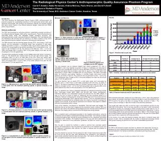
Anthropomorphic Liver Phantom for CT and Ultrasound. By: Katelyn Herbert Advisor: Dr. Bob Galloway. Problem Statement. 20,000 people are diagnosed with liver cancer every year. Hepatocellular Carcinomas (HCCs) Surgical resection only available for 20% of patients. Radiofrequency Ablation.

E N D
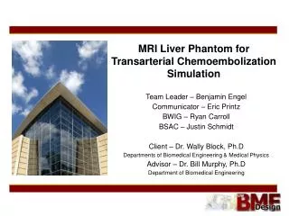
Anthropomorphic Liver Phantom for CT and Ultrasound By: Katelyn Herbert Advisor: Dr. Bob Galloway
Problem Statement • 20,000 people are diagnosed with liver cancer every year. • Hepatocellular Carcinomas (HCCs) • Surgical resection only available for 20% of patients
Radiofrequency Ablation • Needle-like ablation tool is place inside the tumor • High Frequency RF signal heats the surrounding tissue, killing it • Image guided surgery http://www.hopkins-gi.org
Problem Statement • Success of this method is dependent on accurate placement of the tool • 44 percent of all liver tumors are indistinguishable from healthy tissue using this method. • Finding the center of the three-dimensional object (the tumor) in a two-dimensional view is difficult. • Ablation tools made out of flexible materials • placement not always accurate
Objective • Create a phantom that will test the accuracy of the placement of an ablation tool • For training purposes
Importance • Correct placement of an ablation tool insures: • the tumor tissue will be completely removed • Lessen the need for additional surgeries (lessen cost)
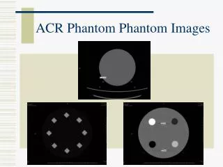
The Model • Anthropomorphic phantom of the liver • Silicone with silicone and barium “tumor” ~ 4 cm • Remove air bubbles from “liver tissue” to allow for use with ultrasound • Create air bubbles in “tumors” to create contrast
The Model 270 mm
The Model • Be able to detect the position of the ablation tool • Send a high frequency signal from a function generator down the ablation tool • Wire around tumors • Signal created in mesh by the presence of the ablation tool • Higher the signal, the closer the tool to the tumor
The Model • Unable to create a signal large enough using this method (LED) • Pairing with antenna and ablation tool less than ideal • Developed new plan of action
The Model Revised • Use of a reed switch Reed Switch www.standexelectronics.com V+ • Reed Switches are unique in that they are triggered by magnetic fields
The Model Revised • Tiny magnet placed in tip of ablation tool • As ablation tool gets within the appropriate distance to the reed switch, it will close and light an LED. kjmagnetics.com
The Model Revised 3.5 mm Reed Switch Ablation Tool Magnetic End Tumor
Progress • Created anthropomorphic phantom out of silicone for use in CT and ultrasound • Removed air bubbles to insure the tumors were visible in the images
Current/Future Work • Test the reed switch with the tiny magnets • Gauge how large of a magnetic field is required to trigger it/ how close it needs to be
Current/Future Work • Insert switch in “tumor” and embed in liver phantom • Test with ablation tool
References • Schöber et al, “Guidance and monitoring of radiofrequency liver tumor ablation with contrast-enhanced ultrasound.”, European Journal of Radiology [0720-048X]yr. 2004 vol. 51 pg. 19 • Meloni et al, “Hepatocellular Carcinoma Treated with Radiofrequency Ablation.” American Journal of Roentgenology 2001 vol. 177 pg. 375 • Gazelle et al. “Tumor Ablation with Radiofrequency Energy”. Radiology 2000 vol. 217 633-646