Download

1 / 39
400 likes | 1.02k Views
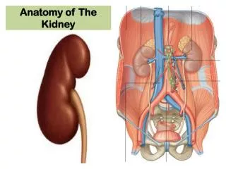
Anatomy, physiology and pathology of the kidney. Dr Andrew Potter Registrar Department of Radiation Oncology Royal Adelaide Hospital. Medical ppt. http://hastaneciyiz.blogspot.com. Anatomy. Overview. Retroperitoneal, paired organs

E N D
Anatomy, physiology and pathology of the kidney Dr Andrew Potter Registrar Department of Radiation Oncology Royal Adelaide Hospital Medical ppt http://hastaneciyiz.blogspot.com
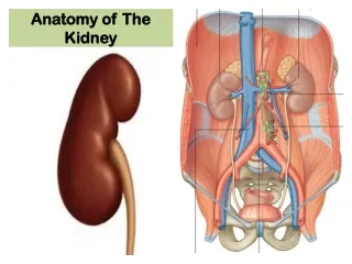
Overview • Retroperitoneal, paired organs • Posterior abdominal wall, largely under cover of costal margin • Key organ of urinary system • Filtration/ concentration of urine • Biochemical balance, hormone production
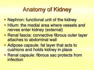
Structure - macro • Enclosed in a strong fibrous capsule which passes over the lips of the sinus and becomes continuous with the walls of the calices. • Kidney + capsule are surrounded by pararenal fat • Each kidney has superior and inferior poles, medial and lateral borders/margins and anterior and posterior surfaces • Reddish-brown in colour when fresh – colour varies between cortex and medulla • Measure ~12x6x3cm (left often slightly longer than right) • Weigh ~130g each • Ovoid in outline but indented medially (the renal sinus) bean-shaped appearance
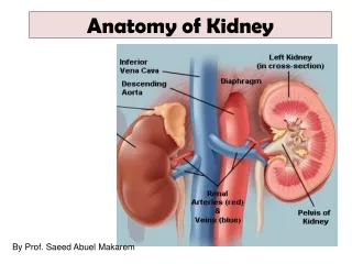
Structure - macro • Hilum • At the concave part of each kidney • Renal vein exits (anteriorly) • Renal artery enters (posterior to renal vein) • Renal pelvis exits (posterior to artery)
Structure - macro • Renal pelvis • Funnel-shaped • Lined with transitional epithelium with a smooth muscle and connective tissue wall • Continuous inferiorly with ureter • Divides into major and minor calyces • Urine collecting tubule minor calyx major calyx renal pelvis ureters bladder
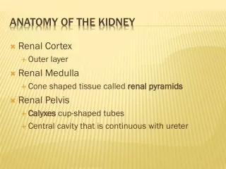
Structure - macro • Cortex • Beneath capsule, extends towards the pelvis as renal columns lying between pyramids of medulla • Apices of several pyramids open together into a renal papilla, each of which projects into a renal calyx
Strcuture - micro • Nephrons • Functional and histological subunit • ~106 per kidney • = glomerulus + tubules • glomerulus • tuft of capillaries surrounded by podocytes • projects into Bowman’s capsule • tubule system • epithelium continuous with Bowman’s capsule • proximal convoluted tubule Loop of Henle distal convoluted tubule collecting tubule and collecting duct • glomeruli and convoluted tubules are in cortex • ducts lie in the medulla • glomerular capillaries supplied by afferent arteriole and drained by efferent arteriole
Position and relations • Lie in a mass of fat (perinephric fat) and fascia, retroperitoneally against posterior abdominal wall • Fatty renal capsule is covered by fibroareaolar tissue – the renal fascia • Renal fascia • encloses kidney, its surrounding fibrous and fatty capsules • helps maintain organ position • superiorly, is continuous with fascia of inferior diaphragm • medially the left and right fascia blend with each other anterior to abdominal aorta and IVC • posterior layer of fascia blends with fascia overlying psoas • Extraperitoneal fat outside the renal fascia is located between peritoneum of posterior abdominal wall and renal fascia
Surface anatomy • Superior poles protected by 11th and 12th ribs • Extend from T12 to L3 vertebral bodies • Move ~2cm superior-inferior during respiration • Right – just below transpyloric plane, 5cm right of midline. Inferior pole ~ finger-width superior to right iliac crest • Left – just above transpyloric plane, 5cm left of midline.
Arterial supply • Renal arteries • branches of aorta at L1/L2 lie behind pancreas and renal veins • Enter at hilum, giving rise to • Anteriorly – apical, upper, middle and lower segments • Posteriorly – posterior segment • No communication between segments
Venous drainage • Renal veins • Communicate widely • Eventually form 56 vessels that unit at the hilum • Drain into IVC
Lymphatic drainage • Para-aortic nodes at L1/L2 • Surface of upper kidney drains through diaphragm into nodes in the posterior mediastinum
Innervation • Sympathetic • Preganglionic cells in spinal cord T12/L1 fibres to thoracic and lumbar splanchnic nerves • Postganglionic cells in coeliac, renal and superior hypogastric plexuses • Vasomotor function
Development • Arises from mesoderm • Pronephros • Transitory, non-functional structures consisting of a few ducts which persist • Mesonephros • Large elongated organs that function as interim kidneys • Glomeruli + tubules open into mesonephric ducts • Metanephros • Permanent kidneys • Begin to develop in ~5th week • Arises caudal to mesonephros • Induces a bud from caudal end of mesonephric duct (ureter) • Ureteric bud divides into calyces of pelvis and collecting tubules and medullary pyramids • Develops in anatomic pelvis and migrates to adult position and the new single definitive artery forms
Physiology - overview • Regulation of the water and electrolyte content of the body • Retention of substances vital to the body such as protein and glucose • Maintenance of acid/base balance • Excretion of waste products, water soluble toxic substances and drugs • Endocrine functions
Water and electrolyte regulation • Renal blood supply is approx 20% of cardiac output • 99% to cortex • 1% to medulla • 2 capillary beds,arranged in series: • Glomerular • High pressure for filtering • Peritubular • Low pressure for absorption
Water and electrolyte regulation • Urine formation - 3 phases • Simple filtration • Selective and passiveresorption • Concentration
Filtration • Takes place through the semipermeable walls of the glomerular capillaries • almost impermeable to proteins and large molecule • Glomerular filtrate is formed by squeezing fluid through glomerular capillary bed • Hydrostatic pressure (head of pressure) is controlled by afferent and efferent arterioles, and provided by arterial pressure • About 20% of renal plasma flow is filtered each minute (125 ml/min). This is the glomerular filtration rate (GFR). • Autoregulation • With a change in arterial blood pressure, there is constriction or dilatation of the afferent and efferent arterioles, the muscular walled vessels leading to and from each glomerulus
Juxtaglomerular apparatus • Macula densa cells • Detect chloride concentration • Juxtaglomerular cells • Modified smooth muscle cells • Produce renin • Converts angiotensin to angiotensin I • Angiotensin I converted to angiotensin II by Angiotensin converting enzyme (ACE) • Causes systemic vasoconstriction and increase in BP
Tubular reabsorption • 60% of solute isreabsorbed inproximal tubule • Different partsof tubule systemoptimised to absorb differentcomponents of urine • Distal tubule and collecting duct determines final urine concentration • Regulated by ADH production by posterior pituitary
Acid-base balance • Tubular acid secretion • Ammonia secreted bytubules (combines withH+ to form NH4+and passed in urine)
Hormones • Renin • Increases production of angiotensin II • Aldosterone • Stimulates water and sodium ion resorption in distal tubule • Atrial natriuretic hormone (ANP) • Produced when atrial pressure increases (eg heart failure) • Promote Na+, Cl- and water loss • Antidiuretic hormone • Increases permability of distal tubule to water, to cinrease water resorption (therfore increases concentration of urine) • 1,25 dihydroxy vitamin D3 • Promotes calcium absorption from gut • Erythropoietin (EPO) • Stimulates marrow to produce red blood cells
Benign pathology • Vascular disease • Hypertension, diabetes, deposition of immune complexes (eg amyloidosis), coagulation • Inflammatory/autoimmune conditions • SLE • Infective • Pyelonephritis, tuberculosis • Idiopathic • Nephrotoxic drugs - eg. platinum chemotherapy, aminoglicoside antibiotics • Congenital/structural • Polycystic kidney, horseshoe kidney, renal agenesis/hypoplasia • Metabolic/biochemical • Renal calculi
Benign tumours • Frequent incidental findings (up to 20%) • Renal adenoma • Bening epithelial tumours arising from tubular epithelium • Difficult to distinguish from renal cell carcinoma - similar histology • Distinguished on size (<3cm)
Benign tumours • Oncocytomas • Variant of adenoma • Angiomyolipoma • Smooth muscle, fat and vessels • Renal fibroma • Common small tumours • 3-10mm • Arise in medulla
Malignant tumours • 90% are renal cell adenocarcinoma (RCC) • About 3% of all adult cancers • Usually seen >50 years of age • Present with haematuria, pain, loin mass • Paraneoplastic syndrome • Hypercalcaemia, hypertension, polycythaemia, Cushing’s syndrome or other hormonal disturbances
Renal cell carcinoma • Rounded masses, yellowish colour with haemorrhage and necrosis • Most commonly the ‘clear cell’ variant • Clear cytoplasm because of high lipid and glycogen content
Renal cell carcinoma • Spread by local extension/expansion through capsule • Blood borne metastases • Bone, lung, brain • Lymphatic metastases • Para-aortic chain • Prognosis depends on stage • 70% ten-year survival of confined to renal capsule • Poor prognosis if metastatic disease at presentation
Nephroblastoma(Wilms’ tumour) • Common childhood malignancy • Embryonal tumour from primitive metanephros • Peak incidence 1-4 years of age • Presents as abdominal mass or haematuria • Rounded mass largely replacing kidney • Solid, fleshy white with necrosis • Prognosis related to stage at presentation
Summary • Paired retroperitoneal/post abdominal organ • Cortex, medulla, nephron • Glomerulus, tubule, duct • Water/biochemical regulation • Filtration, reabsorption • Hormone production • Many benign pathological conditions • Malignancies predominantly RCC in adults, nephroblastoma in children
Medical ppt http://hastaneciyiz.blogspot.com