Download
1 / 22
230 likes | 469 Views
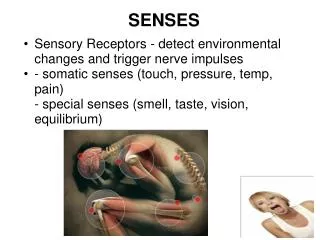
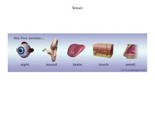
Senses. Sensory relationships. All of our senses respond to stimuli in the environment Each sense has its own specific organ In each sense organ there are specific sense receptors cells

E N D
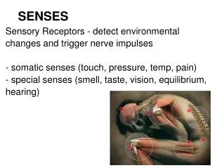
Sensory relationships • All of our senses respond to stimuli in the environment • Each sense has its own specific organ • In each sense organ there are specific sense receptors cells • The role of sense receptors is to register stimuli in the environment and to provide the other parts of the body with information about these stimuli
1. The Eye • The eye is the organ of sight • Our eyes enable us to appreciate: • Lines • colours • light properties • movements of the things around us • The eye is made up of a system of 4 membranes and 3 transparent substances
Anatomy of the Eye Membranes: - 1 membrane covers the front part of the eye the cornea - it is transparent - under the cornea are: 1) The iris : the coloured ring of the eye 2) The pupil: the central portion of this ring (black dot)
Anatomy of the Eye - the other 3 membranes line the posterior (back) of the eye They are all different: • The sclera (outermost membrane) - rigid, gives the eye its shape • The choroid (middle membrane) - provides nourishment for the eye • The retina (innermost membrane) - active nervous membrane = receiver of stimuli
Anatomy of the Eye Transparent Substances: Found inside the eye • The lens • Aqueous humour - located between the cornea and lens - a liquid made up of water and minerals • Vitreous humour - located between the lens and the retina - jelly like substance Eye Intro Video
What path does light take as it travels through the eye? 1) Light must pass through a number of transparent media before it reaches the nerve cells of the eye. Where are these located? On the retina. • Label these different structures in the order that light passes through: vitreous humour, lens, retina, cornea, aqueous humour 1) cornea 2) aqueous humour 3) lens 4) vitreous humour 5) retina
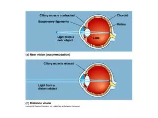
The Path Light Takes in the Eye • Light passes through and is bent by the cornea and then the lens • Images appear inverted on the retina • When looking at near-by objects = the lens curves and thickens • When looking at far object = the lens flattens • The reflex that causes the lens to change shape is called the accommodation reflex • near-far sightedness • The light REFRACTS in the lens; it slows down and changes the direction because of the speed change. Refraction Video
Flattened lens thickened lens Physiology of the Eye Video
Eye Disorders • Hyperopia (far-sighted): • The image is focused behind the retina • Close objects are blurred • Requires CONVEX lenses • Myopia (near-sighted): • The image is focused in front of the retina • Distant objects are blurred • Requires CONCAVE lenses • Presbyopia: • loss of elasticity of the lens (usually in old age) Long eyeball Myopia Short eyeball Hyperopia
Light rays enter the eye and focus on the retinal nerve cells (neurons) • These retinal nerve cells then change the light waves that strike them into nerve impulses that are carried along the optic nerve to the visual center of the brain • Optic nerve • Nerve tissue formed by the axons of the retinal cells • Transmits nerve impulses form the retina to the optical centre of the brain • Examples of retinal nerve cells: • Rods = detect light and dark • Cones = detect colour
What is the role of the brain in vision? • It is only when the center for vision in the brain (in the occipital lobe) is stimulated that a person has any visual sensation • Therefore, it is not only the eyes alone that allow us to “see” but also the brain.
Match the following words with the correct term • Eye • Retina • Optic Nerve • Brain • Transformer • Analyzer • Conductor • Receiver
Recall the Blind-spot? R L Instructions: Close one eye and focus the other on the appropriate letter (R for right or L for left). Place your eye a distance from the screen approximately equal to 3× the distance between the R and the L. Move your eye towards or away from the screen until you see the other letter disappear. For example, close your right eye, look at the "L" with your left eye, and the "R" will disappear.
Normal View Myopia Hyperopia Near Sighted Far Sighted