Download
1 / 77
860 likes | 1.37k Views
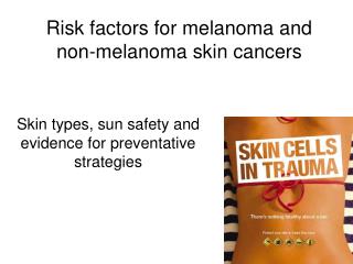
Precancers and Skin Cancers. Adam O. Goldstein, MD, MPH Associate Professor Family Medicine University of North Carolina at Chapel Hill aog@med.unc.edu. Actinic Keratoses. premalignant skin lesions = “keratinocytic intraepidermal neoplasia”

E N D
Precancers and Skin Cancers Adam O. Goldstein, MD, MPH Associate Professor Family Medicine University of North Carolina at Chapel Hill aog@med.unc.edu
Actinic Keratoses • premalignant skin lesions = “keratinocytic intraepidermal neoplasia” • chronic sun, radiation or polycyclic aromatic hydrocarbons • Skin Type I-II • organ transplant
Actinic Keratoses • Distribution: Sunexposed, esp. dorsa hands/forearms • Description: papules,plaques with scale and erythema, occasional crust or cutaneous horn • Sandpapery feel
Actinic Keratoses • epidermal atypia • abnormal maturation
Actinic Keratoses • 60% predisposed >40 have at least 1 AK • 6-10% lifetime >> invasive SCC • >10 AK - 14% an SCC w/n 5 yrs • 60-97% of SCC from AK • ~ 40% of met SCC>> AK • ^ aggressive immsupp
Actinic Keratoses • lip lesions: actinic cheilitis/leukoplakia • white plaques-mucosa • persistent scaling lesions on the lip • ^ aggressive behavior • tobacco/sun
Differential Diagnosis • squamous cell carcinoma: more indurated, thicker, recurrence of AK after treatment
Differential Diagnosis • seborrheic keratosis: hyperpigmented,more stuck on appearing
Differential Diagnosis • nummular eczema: coin-shaped scaling lesions; responds to emollients/topical corticosteroids
AK Treatment • PREVENTION • Screen for skin cancers • Broad-brimmed hats • sun protective clothing • sunscreens • avoidance of sunlight • ed s/sx skin cancer • avoidance of tobacco • low fat diet?
AK Treatment • Cryosurgery(liquid nitrogen) • 5-fluorouracil cream or solution • Diclofenac Sodium-3% gel • Imiquimod 2 x week/ 16 weeks
AK Treatment • Excision • Electrocautery • Curettage • Carbon dioxide laser
AK Treatment • Chemical peels • Photodynamic therapy • Retinoids-topical/oral • Investigational-dimericine
TREATMENT • Liquid Nitrogen-Advantages • cure rates of 98.8% • common • minimal patient ed • multiple/thicker lesions • quick recovery
TREATMENT • Liquid Nitrogen-Disadvantages • storage • pain • pigment alteration • training
5-Fluorouracil • Cure 50-80% • Blocks methylation reaction of deoxyuridylic acid to thymidilic acid • DNA (and RNA) synthesis
Diclonfenac Sodium 3% Topical Gel • mechanism of action unknown • NSAID • inhibition of cyclo-oxygenase >>>PGE-2 • 90 days BID--overall 33-47% clearance vs 10-19% vehicle • avoid ASA triad • hypersensitivity
Photodynamic therapy (Pariser DM - J Am Acad Dermatol -2003)
Cycle therapy of actinic keratoses of the face and scalp with 5% topical imiquimod cream: An open-label trial. Significant irritation Rest periods required Evolving protocols Expensive Effective Salasche SJ et al Am Acad Dermatol 2002;47:571-7.
>1 million cases/yr >50% of all new cancers 1 in 5 Americans will develop skin cancer Skin Cancer Statistics
Types of Skin Cancers • Basal Cell Carcinoma - 80% • Squamous Cell Carcinoma - 16% • Melanoma - 4%
BCC /SCC • Most common skin cancers • Most important risk factors • sun exposure • family history • skin type • Incidence of these cancers increase with age, probably related to cumulative sun exposure
Basal Cell Carcinoma • the most common skin cancer • 90% appear on face, ears, head
Main Types Basal Cell Carcinomas • Nodular BCCs - most common type • Sclerosing BCCs (morpheaform) • Superficial BCCs
Pattern of Nodular BCC • raised pearly white, smooth translucent surface with telangiectasias
Pattern of Nodular BCCs • may ulcerate leaving a small bloody crust • may be pigmented
Pattern of Sclerosing BCCs • ivory or colorless • flat or atrophic • indurated • may resemble scars • are easily overlooked
Pattern of Sclerosing BCCs • ivory or colorless • flat or atrophic • indurated • may resemble scars • are easily overlooked
Pattern of Superficial BCCs and SCC in situ • red or pink scaling plaques • occasionally with shallow erosions or crusts • differentiation between these two similar lesions usually requires a biopsy
Pigmented BCCs • may look like melanoma • increased brown or black pigment • seen more commonly in dark-skinned individuals
Differential Diagnosis of Nodular BCC • Intradermal nevus • Sebaceous hyperplasia • Fibrous papule of the face • trichoepithelioma
Differentiating Intradermal Nevus from Nodular BCC • Intradermal nevus • Stable size • Soft • No crusting or ulceration • May have telangiectasias
Differentiating Intradermal Nevus from Nodular BCC • Intradermal nevus • Stable size • Soft • No crusting or ulceration • May have telangiectasias
Sebaceous Hyperplasia from Nodular BCC • Sebaceous hyperplasia • yellow coloration • stable size • umbilication without ulceration • is hard to see after injecting anesthesia
Diagnosis of Basal Cell Carcinomas • Shave biopsy • nodular • thick superficial types • Punch biopsy • morpheaform • flat superficial types
Treatment options for Basal Cell Carcinomas • C + D after a shave biopsy • Cryotherapy with thermocouple if you have experience • Excision with 3- 5 mm margins • Superficial trunk/ext: imiquimod qd x 12 wks • Mohs for recurrent BCC and areas of cosmetic importance
Mohs micrographic surgery • removal of tumor by scalpel in sequential horizontal layers. • each tissue sample is frozen, stained, and microscopically examined • repeated until all the margins are clear • treatment of choice for BCCs with poorly defined margins • especially those on the nose or eyelids
Recurrence rates after Tx of BCCs • C + D 10% • Cryotherapy 10% • Excision 2 - 5% • Imiquimod ??? • Mohs <1%
Factors that increase recurrence rates • sclerosing vs others • larger size of BCC • margins • experience of the surgeon
Sclerosing BCC is most dangerous • tend to be deeply invasive • often not diagnosed until they have caused extensive damage • invade muscle, nerve, and bone • nodular BCC can also invade deeply
Bowen’s disease - features • SCC in situ • Mainly sun exposed areas • Slightly elevated red scaly plaque with well-demarcated borders
Bowen’s disease - features • May resemble psoriasis, superficial BCC, chronic eczema, SK • Curable using C & D, cryo, 5-FU, imiquimod, excision
Keratoacanthoma • Appear suddenly, grow rapidly • Central crater with keratin plug • May grow to 2cm in size • May resolve spontaneously • May look like SCC
Keratoacanthoma • C and D • elliptical excision • 5-FU topically tid • 5-FU intralesional injection
Location of SCCs • Same distribution as bccs. • Especially on the lips, ears, and scalp • Initially grow by direct extension • Metastasize to local lymph nodes and then to distant sites
SCCs with an increased risk of metastasis • larger, advanced lesions • SCC on mucous membranes (in the oral cavity, on the lips) • BCCs rarely metastasize
SCC more aggressive (local & mets) • Size >2 cm • SCC in a scar • Patient is immunosuppressed • Poorly differentiated • There is perineural invasion
Importance of early diagnosis of BCC and SCC • especially in facial cancers • the nose is the single most frequent site of BCC • reconstruction is difficult • extension into underlying bone and cartilage may occur
The differential diagnosis of superficial BCC and SCC in situ • Actinic keratosis, nummular eczema • Nummular eczema can usually be distinguished by its coin-like shape, transient nature, and itchiness • Biopsy any thickened and crusting actinic keratosis to rule out BCC or SCC