Download
1 / 5
60 likes | 469 Views
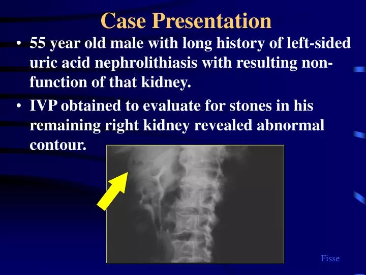
Case Presentation. 55 year old male with long history of left-sided uric acid nephrolithiasis with resulting non-function of that kidney. IVP obtained to evaluate for stones in his remaining right kidney revealed abnormal contour. Fisse.

E N D
Case Presentation • 55 year old male with long history of left-sided uric acid nephrolithiasis with resulting non-function of that kidney. • IVP obtained to evaluate for stones in his remaining right kidney revealed abnormal contour. Fisse
Ultrasound reveals a solid mass near the lower of the two collecting systems. • The left image is shows the typical ultrasound appearance of a duplicated collecting system with hypoechoic parenchyma separating the hyperechoic peripelvic fat and vessels into upper and lower moieties Fisse
Renal scan shows too little function in left kidney to allow nephrectomy without rendering patient dialysis dependant Repositioning of “region of interest” shows lower pole containing tumor has a fair percentage of his renal function making heminephrectomy also a less attractive option 12% 57% 43% 88% Fisse
Partial nephrectomy performed Intraoperative View Specimen (Renal Cell Carcinoma) Fisse