Download
1 / 57
570 likes | 833 Views
Carcinoma of the Vulva Epidemiology. 4% of gynecologic malignancy The median age of patients with invasive vulvar cancer at diagnosis is about 65 to 70 years median age of women with VIN at diagnosis is 45 to 50 years.

E N D
Carcinoma of the VulvaEpidemiology • 4% of gynecologic malignancy • The median age of patients with invasive vulvar cancer at diagnosis is about 65 to 70 years • median age of women with VIN at diagnosis is 45 to 50 years
The relatively stable incidence of invasive cancer despite a steady increase in patients diagnosed with VIN could suggest • etiologic factors are different • Dx improved • effective treatment of VIN has prevented a significant increase in the incidence of invasive disease.
HPV • 89 percent of VIN lesions • 40% of invasive vulvar carcinomas • HPV vaccination
Invasive SCC • associated with HPV infection • notassociated with HPV infection
HPV-positive tumors • Basaloid or warty carcinomas with little keratin formation • often associated with VIN • frequently multifocal • in younger women (35 to 55 years) • more likely to have CIN • to have risk factors typically associated with cervical cancer
HPV-negative tumors • In older women (55 to 85 years) • often associated with vulvar inflammation or lichen sclerosis (but rarely with VIN) • Unifocal • well differentiated • Higher incidence of p53 mutations
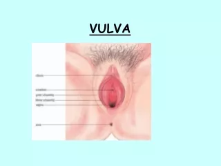
female external genitalia • mons pubis, • labia majora, labia minora • clitoris, • vestibular bulb, • vestibular glands (including Bartholin's glands • vestibule of the vagina.
gynecologic perineum • The region between the posterior commissure of the labia and the anus is termed the gynecologic perineum.
lymphatics Even minimally invasive • superficial inguinal lymph nodes(lateralized lesions) • deeper femoral lymph nodes (secondarily ) • pelvic lymph nodes (then) • medial femoral lymph nodes(more medial lesions) • obturator nodes(clitoris)
Despite the extensive anastomosis of lymphatics in the region, metastasis of vulvar carcinoma to contralateral lymph nodes is uncommon in patients with well-lateralized T1 lesions.
The lungs are the most common sites of hematogenous metastasis
Pathology Nonneoplastic epithelial disorders of the vulva • lichen sclerosis • squamous hyperplasia • dermatoses
About 10% of these lesions have cellular atypia and are termed vulvar intraepithelial neoplasia • VIN lesions are assigned a grade from 1 to 3 according to their degree of maturation
Paget's disease of the vulva • a rare intraepithelial lesion located in the epidermis and skin adnexa, accounts for 1% to 5% of vulvar neoplasms. • negative for HPV • in postmenopausal women
SCC(More than 90% ) Most squamous carcinomas are well differentiated, About 5% of vulvar cancers are anaplastic carcinomas
Verrucous carcinoma • Rare • very well-differentiated • in the fifth or sixth decade • a large, locally invasive lesion. • Even with extensive local invasion, lymph node metastasis from verrucous carcinoma is very rare.
primary mammary adenocarcinoma • basal cell carcinomas • sebaceous carcinomas • Malignant melanomas • Vulvar sarcomas
Diagnosis, Clinical Evaluation, and Staging Patients with VIN may complain of : • vulvar pruritus • irritation, • a mass • 50% are asymptomatic • bleeding • tender
new vulvar lesion • biopsy • Once the diagnosis of high-grade VIN has been established, the entire vulva, cervix, and vagina should be carefully examined because patients often have multifocal or multicentric involvement.
Colposcopic examination • wedge biopsy • Excisional biopsy is preferred for lesions smaller than 1 cm in diameter.
a careful physical examination • chest radiography • biochemical profile • Cystoscopy and proctoscopy • skeletal radiography • CT or MRI scans • PET (poor sensitivity but high specificity in the prediction of lymph node metastases)
(FIGO) Staging of Carcinoma of the Vulva • I Lesions 2 cm or less in size confined to the vulva or perineum. (T1) (N0) • IA Lesions 2 cm or less in size confined to the vulva or perineum and with stromal invasion no greater than 1.0 mm (No) • IB Lesions 2 cm or less in size confined to the vulva or perineum and with stromal invasion greater than 1.0 mm (No)
II Tumor confined to the vulva and/or perineum or more than 2 cm in the greatest dimension. (T2) (N0) • III Tumor of any size with:Adjacent spread to the lower urethra and/or the vagina, or the anus (T3) and/or Unilateral regional node metastasis (N1)
IVA Tumor invades any of the following: upper urethra, bladder mucosa, rectal mucosa, pelvic bone (T4) and/or Bilateral regional node metastasis. (N2) • IVB Any distant metastasis including pelvic lymph nodes. (M1)
Px • Clinical tumor diameter • depth of invasion • tumor thickness • presence or absence of LVSI • tumor grade? • More than 75% of patients with LVSI have positive inguinal nodes.
Prognostic Factors • amount of keratin • the mitotic rate • the tumor growth pattern • Aneuploid tumors (not be an independent predictor of outcome) • HPV DNA ( a poorer prognosis) • age ?
LN • presence and number of inguinal node metastases • bilateral node involvement • pelvic node metastases (as stage IV) • extracapsular extension
surgical margins and tumor recurrence 1 cm or <8mm
Treatment • Radical en bloc resection of the vulva, and inguinofemoral nodes until the early 1980s. • 5-year survival rates of 60% to 70%, • the surgery caused significant physical and psychological complications, • patients with multiple positive nodes continued to have a poor prognosis.
operating through separate vulvar and groin incisions • cure rates similar to vulvectomy. • role of radiotherapy in the curative management of locoregionally advanced disease.
Preinvasive Disease (VIN)- • treatment of high-grade VIN (VIN 3) should be as conservative as possible • Focal lesions can be simply excised. • Multiple lesions can be excised separately or, if confluent, with a larger single excision. • This approach is generally well tolerated and provides material for histologic assessment. • more extensive high-grade VIN, with a CO2 laser.
Extensive, diffuse VIN 3 • a wider excision, particularly if the lesion involves the perianal skin. • a partial vulvectomy of the superficial skin (skinning vulvectomy)
VIN 3 often recurs • VIN 3 can recur within the donor skin from split-thickness grafts
T1 and T2 Tumors • Invasive vulvar tumors can usually be treated effectively without en bloc radical vulvectomy and inguinal node dissection. • Today, most gynecologic oncologists advocate an individualized approach to early invasive vulvar carcinomas.
Overall 5-year disease-specific survival rates for stage I (T1N0M0) and stage II (T2N0M0) disease are approximately 98% and 85%, respectively.
T1 and selected T2 lesions • radical local excision. • A wide and deep excision of the lesion is performed, with the incision extended down to the inferior fascia of the urogenital diaphragm. • An effort should be made to remove the lesion with a 1-cm margin of normal tissue in all directions unless this would require compromise of the anus or urethra.
Small T1 lesions that invade 1 mm or less can be managed with local resection alone because the risk of regional spread is very small.
Larger T2 lesions : modified radical or radical vulvectomy • separate vulvar and groin incisions.
Acute complication • Wound seroma (15% ) • urinary tract infection • wound cellulitis • temporary anterior thigh anesthesia from femoral nerve injury • thrombophlebitis • pulmonary embolus.
Chronic complication • leg edema • genital prolapse • urinary stress incontinence • temporary weakness of the quadriceps muscle • introital stenosis. • pubic osteomyelitis, • femoral hernia, • rectoperineal fistula
T3 and T4 Tumors • T3 tumors S +_RT • the vulva may be treated with opposed anterior and posterior photon fields (if the inguinal regions also require treatment) or with an appositional perineal electron beam. The vulva should receive a total dose of 50 to 65 Gy, depending on the proximity of disease to the surgical margin.
preoperative chemoradiation in some patients with T3 and T4 • These reports indicated that modest doses of radiation (45 to 55 Gy) produced dramatic tumor responses in some patients with T3 and T4 disease, permitting organ-sparing surgery without sacrifice of tumor control. • Investigators have emphasized the use of concurrent chemoradiation in this setting.
Chemoradiation in Locally Advanced Disease • Most studies have used combinations of cisplatin ,5-FU, and mitomycin-C, • Treatment schedules usually include a 4- to 5-day infusion of 5-FU combined with one of the other two drugs, with this course repeated every 3 to 4 weeks
CHRT • Impressive responses • Cisplatin
neoadjuvant chemotherapy in the treatment of locally advanced vulvar cancer. • two to three cycles of cisplatin, bleomycin, and methotrexate followed by radical surgery. • Caution is warranted Elderly (concurrent medical problems)
Tx: • Small T1 lesions : local resection alone • T1 and selected T2 lesions: radical local excision • Larger T2 lesions: modified radical or radical vulvectomy • Locally Advanced Disease: CHRT +S T3 : S+_RT T4 Tumors:RT+_S+_CHT
Treatment of Regional Disease • patients who suffer inguinal recurrences are rarely curable • primary tumors that invade more than 1 mm must have their inguinal nodes treated
compared pelvic lymphadenectomy with inguinal and pelvic irradiation in patients with inguinal node metastases from carcinoma of the vulva: a survival advantage for the radiotherapy arm
postoperative radiotherapy became standard for most patients with inguinal node metastases.